Psilocybin & Neuroscience
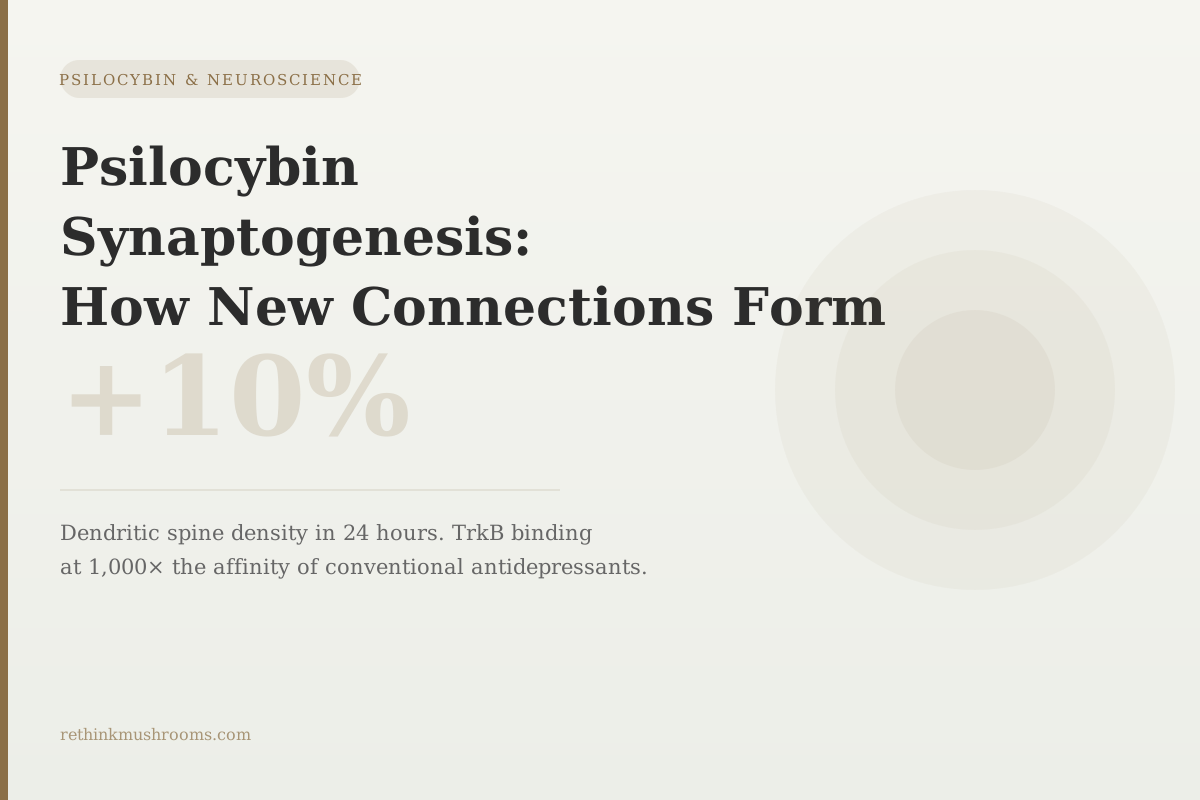
10%
A single psilocybin dose increased dendritic spine density — the structural basis of synapses — by approximately 10% in mouse frontal cortex within 24 hours, with new spines persisting for at least one month.
Shao LX et al., Neuron, 2021
1,000×
higher affinity for the TrkB growth receptor than conventional antidepressants (Moliner et al., Nature Neuroscience, 2023)
24 hours
onset of dendritic spine growth in mouse frontal cortex after psilocybin (Shao 2021)
1 month
persistence of new dendritic spines after a single psilocybin dose (Shao 2021)
Shao LX et al., Neuron, 2021
 View as image ↗
Every time the brain learns something new, it physically changes: neurons grow new connection points, called synapses, that allow cells to communicate more efficiently along pathways related to the new information. This process — synaptogenesis, the formation of new synapses — is what makes the brain adaptive. Without it, learning, memory, and recovery from psychological injury would be impossible.
Psilocybin promotes synaptogenesis. The evidence for this comes from four studies using different methods: cell cultures, mouse brain imaging, pig brain imaging, and molecular binding experiments. Together they identify three distinct molecular pathways through which psilocybin triggers new synapse formation, explain why structural changes persist long after the drug clears, and show a 2023 paradigm shift that reframes how the whole mechanism works.
All of this evidence is preclinical — mice, pigs, and cell cultures. No study has directly measured synaptogenesis in a living human brain after psilocybin. Understanding what the evidence does and does not show is as important as understanding the findings themselves. For the broader neuroscience picture of how psilocybin affects brain function, including Default Mode Network effects, see psilocybin and the Default Mode Network: how it quiets the ego.
Key Takeaways
- Ly et al. (2018, Cell Reports) established that psilocybin and other classic psychedelics promote synaptogenesis through three molecular pathways: activation of TrkB (the brain's growth receptor), activation of mTOR (the cell's growth regulation master switch), and 5-HT2A receptor signaling.
- Moliner et al. (2023, Nature Neuroscience) discovered that psilocin directly binds TrkB with affinities approximately 1,000-fold higher than conventional antidepressants — and that the neuroplasticity and antidepressant effects in mice depend on this TrkB binding, not on 5-HT2A activation.
- Shao et al. (2021, Neuron) used two-photon microscopy (real-time imaging through a window in the mouse skull) to show a single psilocybin dose grew new dendritic spines — the protrusions that form the receiving ends of synapses — by approximately 10% in 24 hours, with changes persisting one month.
- Raval et al. (2021, Neuropsychopharmacology) provided the most translational evidence by using PET imaging in pig brains: a single psilocybin dose measurably increased synaptic density, as detected by SV2A (a protein found exclusively in synaptic vesicles), across multiple brain regions.
- The Moliner 2023 finding separates two previously linked mechanisms: the subjective psychedelic experience (driven by 5-HT2A activation) and the structural neuroplasticity (driven by TrkB binding). These are independent effects.
- All synaptogenesis evidence is preclinical; no direct imaging of synaptic density changes after psilocybin exists in humans yet.
Synaptogenesis vs. Neurogenesis: The Difference That Matters
Before getting into the evidence, it helps to clarify what synaptogenesis is and what it is not.
Synaptogenesis is the formation of new synapses between existing neurons. A synapse is the physical connection point where one neuron sends a chemical signal to another. More synapses along a given pathway means that pathway communicates more efficiently and reliably. The formation of new synapses — synaptogenesis — is what underlies learning, adaptation, and recovery.
Synaptogenesis is not the same as neurogenesis, which is the growth of entirely new neurons. Neurogenesis also occurs in the adult brain, primarily in the hippocampus (the brain's memory formation center), but it is a slower and more restricted process. When people say psilocybin "grows new brain cells," they are usually describing the wrong process. The synaptic growth evidence is about new connections forming on neurons that already exist.
The structures involved are dendritic spines. A neuron's dendrites are the branching extensions that receive incoming signals. Along these branches, tiny protrusions called dendritic spines (each about 1 micrometer in diameter) form the receiving end of individual synapses. A single neuron can have thousands of dendritic spines. More spines mean more potential communication channels with other neurons. Synaptogenesis, in structural terms, is the growth of new dendritic spines and the formation of synaptic contacts on them.
Depression has a synaptic component: major depressive disorder is associated with reduced dendritic spine density in the prefrontal cortex (the brain's planning and self-regulation center), driven by chronic stress and elevated stress hormones that accelerate synaptic pruning. The therapeutic relevance of psilocybin-induced synaptogenesis is that it may reverse this pruning.
Three Molecular Pathways: How Psilocybin Triggers New Synapses
The Ly et al. 2018 paper, published in Cell Reports from David Olson's lab at UC Davis, established the scientific framework for understanding psychedelics as promoters of neural plasticity.[1] The paper introduced the term "psychoplastogens" — compounds that rapidly and robustly promote structural and functional plasticity — and demonstrated this effect across multiple classic psychedelics including psilocybin, LSD, and DMT.
The study identified three molecular pathways through which psychoplastogens trigger synaptogenesis:
TrkB pathway. TrkB (tropomyosin receptor kinase B) is the primary receptor for BDNF — brain-derived neurotrophic factor, the brain's most important growth and survival protein. When TrkB is activated, it initiates signaling cascades that promote neurite growth (the extension of neuronal branches), spinogenesis (new spine formation), and synaptogenesis. The Ly 2018 study showed that blocking TrkB significantly reduced the plasticity-promoting effects of psychoplastogens.
mTOR pathway. mTOR (mechanistic target of rapamycin) is the cell's master regulator of growth and protein synthesis — the central switch that decides when a cell should grow new structures. Psychedelic activation of mTOR drives the cellular machinery required to physically build new spines and synaptic contacts. Blocking mTOR also reduced the plasticity effects.
5-HT2A pathway. The 5-HT2A receptor — the primary target of psilocybin's hallucinogenic effects — is also involved in synaptogenesis signaling. Activation of 5-HT2A initiates downstream pathways that intersect with the TrkB and mTOR cascades. For an in-depth look at how 5-HT2A activation produces the subjective psychedelic experience, see how 5-HT2A agonism creates the psychedelic experience.
What the Ly 2018 study showed is that all three pathways contribute, and they do so synergistically — blocking any one reduces but does not eliminate the plasticity effect. This redundancy may explain why psychedelic-induced synaptogenesis is so robust compared to other interventions.
The 2023 TrkB Discovery That Reframed the Whole Story
The Moliner et al. 2023 paper, published in Nature Neuroscience from the Castrén lab, is the biggest mechanistic update in this field since Ly 2018.[2] It changed the fundamental understanding of how psychedelics produce neuroplasticity.
The discovery: psilocin (the active form of psilocybin after it crosses the blood-brain barrier) directly binds to TrkB. Not indirectly, through BDNF or serotonin signaling cascades, but through direct molecular binding. The binding site is in the transmembrane domain of TrkB — the region that spans the cell membrane — rather than the extracellular domain where BDNF binds.
The affinity of this binding is approximately 1,000-fold higher than the TrkB binding affinity of conventional antidepressants, including SSRIs and SNRIs, which also activate TrkB but only weakly. To put that in concrete terms: psilocin activates the brain's growth receptor far more powerfully than any currently approved antidepressant.
The implication that Moliner pursued experimentally was whether this TrkB binding is actually necessary for the neuroplasticity and antidepressant effects — or whether it is just an incidental molecular interaction. They found it is necessary. In mice where TrkB binding was prevented (by using a mutant form of TrkB that blocks this specific interaction), psilocybin's neuroplasticity effects and antidepressant-like behavioral effects were significantly reduced, even though 5-HT2A was still being activated.
This separates two things that were previously assumed to go together:
The subjective psychedelic experience is driven by 5-HT2A activation. The structural neuroplasticity is driven by TrkB binding. They are parallel mechanisms that happen to co-occur when psilocybin is present — because psilocin activates both receptors — but they can, in principle, be separated.
This is why the Moliner 2023 finding generated significant interest: if the neuroplasticity benefit comes from TrkB binding, it might be possible to develop compounds that bind TrkB with high affinity without also activating 5-HT2A — producing the therapeutic structural benefit without the psychedelic experience. That research line is now being pursued.
Watching New Synapses Form in Real Time: The Shao 2021 Data
While Ly 2018 and Moliner 2023 explain the molecular pathway, the Shao et al. 2021 paper from Yale, published in Neuron, provided direct visual evidence of the structural changes.[3]
The experimental method was two-photon microscopy — a laser imaging technique capable of creating real-time images of individual cells inside living brain tissue without damaging the tissue. To use this technique, researchers implanted a tiny transparent window in the skull of mice, allowing the microscope to peer into the living cortex. Over multiple imaging sessions before and after psilocybin administration, they could directly track whether individual dendritic spines appeared, disappeared, or changed size.
The finding: a single psilocybin dose produced approximately a 10% increase in dendritic spine density in layer 5 pyramidal neurons (the primary output cells) of the mouse medial frontal cortex within 24 hours. The new spines were also larger on average — a larger spine indicates a stronger, more established synaptic connection.
When the researchers came back to image the same neurons one month after the psilocybin dose, the new spines were still there. Structural changes that formed within 24 hours persisted for at least a month — a period during which the drug itself had long been metabolized and eliminated.
The frontal cortex location matters. The medial frontal cortex in mice is broadly equivalent to the prefrontal cortex in humans — the brain region responsible for executive function, emotional regulation, and the kind of top-down control over behavior that becomes impaired in depression and anxiety. This is also the region where synaptic pruning driven by chronic stress is most apparent. The Shao finding that psilocybin grows new spines specifically in this region is consistent with the therapeutic hypothesis.
For a deeper look at the Shao 2021 study specifically — including how the two-photon method works and what the spine persistence data means — see psilocybin and dendritic spine growth: structural neuroplasticity in 24 hours.
From Mice to Larger Brains: PET Imaging in Pigs
The mouse and cell-culture studies establish the mechanism. The pig brain PET study by Raval et al. 2021, published in Neuropsychopharmacology, provides the most translational evidence in the preclinical literature.[4]
The method uses PET (positron emission tomography) imaging with a radiotracer that binds to SV2A — a protein found exclusively inside synaptic vesicles, the small packets that store and release neurotransmitters at synapses. Because SV2A is present in every synapse but nowhere else, its density measured by PET is a reliable proxy for total synaptic density across the brain. This same SV2A-PET methodology is used in human studies of synaptic density changes in depression and other conditions.
Raval found that a single psilocybin dose increased SV2A binding across multiple pig brain regions — meaning measurably more synapses were present after treatment than before. The pig brain data matters because pig brains are larger, more structurally complex, and more similar to human brains than mouse brains. It is the closest existing evidence to what might occur in humans.
The same study also showed a reduction in 5-HT2A receptor density after psilocybin — the expected compensatory downregulation when 5-HT2A is activated. This is consistent with the Moliner 2023 finding that TrkB (not 5-HT2A) is the driver of sustained plasticity: the 5-HT2A receptors are transiently occupied and then downregulated, but TrkB-mediated structural changes persist.
For the human side of this picture — what fMRI and functional connectivity studies show about how psilocybin changes brain network communication — see functional connectivity changes under psilocybin: fMRI evidence. Structural synaptogenesis and functional connectivity changes are complementary aspects of how psilocybin reorganizes brain circuitry.
The Preclinical Wall: What We Don't Yet Know in Humans
Everything described in this article — the three pathways, the 10% spine increase, the 1,000-fold TrkB binding affinity, the PET evidence in pigs — is preclinical evidence. None of it comes from measuring human brain tissue directly.
Human studies of psilocybin's effects on the brain rely on fMRI (which measures blood flow patterns, not synapses), EEG (which measures electrical activity, not synaptic density), or behavioral and clinical measures (which are outcomes, not mechanisms). These methods tell us that psilocybin changes how brains function and that people feel better after treatment. They cannot directly measure whether new synapses form.
The most direct path to human synaptogenesis data would be SV2A-PET imaging of psilocybin-treated patients before and after a session — using the same methodology Raval 2021 used in pigs. This approach has been used to detect synaptic density reduction in depression and schizophrenia using human participants. It has not yet, as of 2025, been applied to psilocybin treatment in humans.
The therapeutic plausibility of the synaptogenesis hypothesis in humans rests on three points. First, the molecular mechanisms (TrkB, mTOR, BDNF) are conserved across species — they work the same way in mice, pigs, and humans at the cellular level. Second, the clinical outcomes (remission of depression, lasting changes after a single session) are consistent with what you would expect if structural synaptogenesis were occurring. Third, the PET pig data uses human-compatible methodology and produces consistent results.
Consistent is not proof. The honest position is that the preclinical evidence for psilocybin-induced synaptogenesis is strong and mechanistically well-characterized, and the clinical evidence is consistent with it, but direct human confirmation awaits the right study.
For more on the broader neuroplasticity picture — how synaptogenesis relates to the plasticity window and what the timing of structural changes means for therapy — see the plasticity window: how long psilocybin keeps the brain flexible and psilocybin and neuroplasticity: rewiring the brain.
The Bottom Line
Psilocybin promotes synaptogenesis through three molecular pathways (TrkB, mTOR, and 5-HT2A) identified in the Ly 2018 Cell Reports paper. The 2023 Moliner Nature Neuroscience study added a paradigm shift: psilocin directly binds TrkB at 1,000-fold higher affinity than conventional antidepressants, and this TrkB binding — not 5-HT2A activation — drives lasting structural neuroplasticity. Shao 2021 (Neuron) directly observed ~10% dendritic spine growth in mouse frontal cortex within 24 hours, persisting one month. Raval 2021 (Neuropsychopharmacology) showed synaptic density increases via SV2A-PET in pig brains. All evidence is preclinical; direct human synaptogenesis data does not yet exist.
Frequently Asked Questions
Sources & References
- 1RTHM-00983·Ly, Calvin et al. (2018). “Psychedelics Promote Structural and Functional Neural Plasticity..” Cell reports.Study breakdown →PubMed →↩
- 2RTHM-01756·Moliner, Rafael et al. (2023). “Psychedelics promote plasticity by directly binding to BDNF receptor TrkB..” Nature neuroscience.Study breakdown →PubMed →↩
- 3RTHM-01434·Shao, Ling-Xiao et al. (2021). “Psilocybin induces rapid and persistent growth of dendritic spines in frontal cortex in vivo..” Neuron.Study breakdown →PubMed →↩
- 4. Source details for this reference are not yet available.