Meshima & Cancer
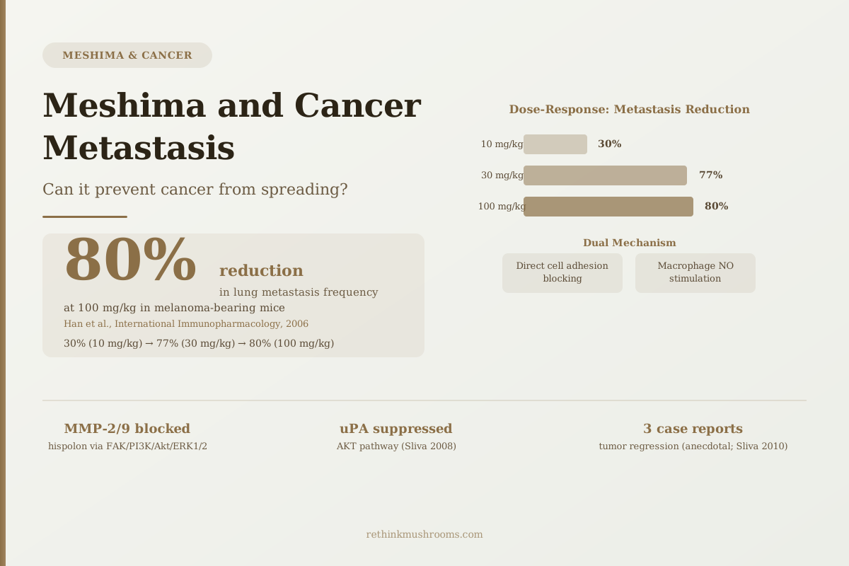
80% reduction
Phellinus linteus polysaccharides reduced lung metastasis frequency by 80% in melanoma-bearing mice at 100 mg/kg — through dual mechanisms: direct cancer cell adhesion blocking and macrophage immune activation.
Han et al., International Immunopharmacology, 2006
30% at 10 mg/kg
dose-dependent — even the lowest test dose produced significant metastasis reduction
77% at 30 mg/kg
intermediate dose; near-maximum effect achieved before top dose
3 case reports
tumor regression in advanced cancer patients from the 2010 Sliva review
Han et al., International Immunopharmacology, 2006
 View as image ↗
Most discussions of anti-cancer compounds focus on tumor killing — shrinking tumors, inducing cell death. The harder problem in cancer biology is metastasis: the process by which cancer cells break away from the original tumor, travel through the bloodstream or lymphatic system, and colonize new organs. Most cancer deaths result not from the primary tumor but from this spread.
Meshima (Phellinus linteus) has been studied specifically for its effects on this process. The research doesn't just show that it kills cancer cells in a dish — it shows that polysaccharide extracts reduce the frequency of new metastatic colonies forming in lungs, block the molecular machinery cancer cells use to invade tissue, and activate immune responses that intercept circulating tumor cells. The most striking single finding: an 80% reduction in lung metastasis frequency in animal models.
All of this data is preclinical. No clinical trial has tested Meshima specifically for its anti-metastatic effects in cancer patients. The evidence is important, mechanistically specific, and nowhere close to establishing clinical efficacy. Both things are true.
For Meshima's general research history and why it's been studied in Japan and Korea for decades, see what Meshima mushroom is and why it's called Korea's anti-cancer mushroom.
Key Takeaways
- Han et al. (2006) showed that Phellinus linteus acidic polysaccharides reduced lung metastasis frequency in melanoma-bearing mice by 30%, 77%, and 80% at doses of 10, 30, and 100 mg/kg respectively
- The anti-metastatic mechanism in Han's study was dual: direct inhibition of cancer cell adhesion to the extracellular matrix AND immunopotentiation through increased macrophage nitric oxide production
- Hispolon, Meshima's key polyphenol, blocks matrix metalloproteinases MMP-2 and MMP-9 — the enzymes cancer cells use to dissolve tissue barriers — through FAK/PI3K/Akt and ERK1/2 pathways (Huang 2010)
- Sliva et al. (2008) showed Meshima suppresses urokinase-plasminogen activator (uPA) secretion in invasive breast cancer cells — blocking a second invasion enzyme independently of MMP inhibition
- Song et al. (2011) demonstrated MMP-9 inhibition in colon cancer cells with reduction of Wnt/β-catenin signaling, plus in vivo tumor reduction in nude mouse xenografts
- The Sliva 2010 review includes 3 case reports of tumor regression in advanced cancer patients — the only published human-adjacent data, and it is anecdotal
The 80% Metastasis Reduction: What Han's 2006 Study Actually Measured
Han and colleagues published what remains the most compelling quantitative anti-metastasis finding for any Meshima compound in 2006 in International Immunopharmacology.[1]
They isolated acidic polysaccharides from Phellinus linteus and tested three doses in B16F10 melanoma-bearing mice: 10, 30, and 100 mg/kg. The endpoint was lung metastasis frequency — how often new tumor colonies formed in the lungs, the primary target organ for melanoma metastasis.
The dose-response was striking. At 10 mg/kg, lung metastasis frequency dropped by 30% compared to controls. At 30 mg/kg, it dropped by 77%. At 100 mg/kg, the reduction reached 80%.
Several things about this finding deserve emphasis. The reduction was dose-dependent and consistent across the three dose levels — not a single dramatic data point. The effect began at the lowest dose tested, which matters because it suggests the mechanism is engaged even at sub-maximal doses. And the mechanism was identified: it was not primarily cytotoxic. The polysaccharides were not killing melanoma cells directly. They were blocking the specific behaviors cancer cells use to establish metastatic colonies.
An earlier study from the same research group (Han et al., 1999) established additional context: PL polysaccharides prolonged survival in melanoma-bearing mice, inhibited tumor growth in NCI-H23 nude mice, and the combination of PL with adriamycin (doxorubicin) was synergistic for tumor growth inhibition.[2] The immune mechanism was also identified in the 1999 study, consistent with what the 2006 paper confirmed.
Two Mechanisms in One Study: Direct Cell Blocking and Immune Activation
The mechanistic finding in Han's 2006 study is what distinguishes it from most anti-cancer mushroom research. The researchers identified two independent pathways operating simultaneously.
The first is direct: PL polysaccharides physically interfere with cancer cell adhesion to the extracellular matrix. Metastasis requires cancer cells to grab onto tissue surfaces before they can colonize them. If adhesion is blocked, the circulating tumor cells cannot attach and establish new colonies. Han's group showed reduced adhesion in cell assays alongside the in vivo metastasis reduction, confirming this as a contributing mechanism.
The second is immunological: PL polysaccharides increased macrophage production of nitric oxide. Macrophages are immune cells that patrol tissues and can kill abnormal cells. Nitric oxide is one of their primary killing tools. By stimulating higher NO production, PL-treated macrophages became more aggressive in targeting cancer cells that do successfully arrive at distant sites.
The combination of these two mechanisms — less cancer cell adhesion at the destination, and a more hostile immune environment when cells do arrive — represents a comprehensive anti-metastatic strategy. Notably, the paper confirmed no direct cytotoxicity on the cancer cells themselves. This is mechanistically important: the polysaccharides aren't working by killing tumor cells outright. They're working by disrupting the process of spread specifically.
The polysaccharide mechanisms that make this possible are described in more detail in Phellinus linteus bioactive compounds: polysaccharides and phenols, which covers how PL polysaccharide structure relates to biological activity.
How Hispolon Dismantles the Molecular Tools Cancer Uses to Invade
While the polysaccharide data addresses the systemic metastasis picture, hispolon — Meshima's phenolic compound — works at a more granular level: blocking the specific enzymes cancer cells use to dissolve tissue and create pathways for movement.
Huang and colleagues tested hispolon against SK-Hep1 human hepatoma cells in 2010.[5] Matrix metalloproteinases (MMPs) are enzymes that break down the structural proteins forming the barriers between cell layers — if cancer cells cannot dissolve these barriers, they cannot invade. Hispolon decreased both MMP-2 and MMP-9 expression in a concentration-dependent manner.
The pathways involved: hispolon suppressed FAK signaling, which sits upstream of PI3K/Akt, RhoA, and ERK1/2 — the signaling cascade that cancer cells use to produce MMP-2 and MMP-9. Testing an ERK1/2 inhibitor (PD98256) also decreased MMP-2 and MMP-9, confirming that ERK1/2 is a critical node in the pathway. The study also showed reduced uPA activity alongside the MMP suppression.
Separately, Sliva and colleagues showed in their 2008 British Journal of Cancer study on MDA-MB-231 breast cancer cells that PL suppressed uPA secretion through AKT pathway inhibition — a mechanistically independent route to blocking the same invasion process.[3] uPA (urokinase-plasminogen activator) is a different class of enzyme from MMPs but serves the same function in enabling cancer cell invasion. Meshima's hispolon component blocks MMPs; the polysaccharide/whole-extract component blocks uPA. Two molecular tools of invasion, blocked by different Meshima compounds through different pathways.
Lu and colleagues also found that hispolon inhibited proliferation in both breast and bladder cancer cells via MDM2-recruited ERK1/2 activity — establishing hispolon as a broad anti-proliferative compound beyond its invasion-blocking mechanisms.[8]
For a detailed look at how Meshima specifically blocks cancer cell migration through EMT markers and cytoskeletal pathways, see Phellinus linteus and cancer cell migration inhibition.
Colon Cancer, Wnt/β-Catenin, and an In Vivo Xenograft
Song and colleagues published a particularly detailed study in BMC Cancer in 2011 using SW480 human colon cancer cells and protein-bound polysaccharide from Phellinus linteus.[4]
At concentrations of 125 to 1,000 μg/mL, PL polysaccharide significantly inhibited SW480 cell proliferation. It decreased expression of β-catenin and cyclin D1, and inhibited both MMP-9 activity and cell motility. The Wnt/β-catenin pathway — which drives proliferation and is dysregulated in approximately 90% of colorectal cancers — was specifically disrupted.
The study went beyond cell culture. The researchers implanted SW480 cells into nude mice and tested whether PL treatment reduced tumor growth. Tumor growth was significantly decreased in the PL-treated animals. The apoptotic index — a measure of how many tumor cells were actively dying — increased in tumor tissue. β-catenin expression was also decreased in the tumor tissue itself, confirming that the Wnt/β-catenin disruption seen in cell culture was occurring in the living tumor.
This is the closest published data to a colon-cancer-specific anti-metastatic in vivo model for Meshima. It doesn't measure metastasis directly — it measures tumor growth inhibition in a xenograft model — but the MMP-9 inhibition finding connects to the metastasis mechanism via the invasion cascade.
A 2023 systematic review of PL polysaccharide structure and function by Qin and colleagues confirmed that anti-tumor, immunomodulatory, and anti-inflammatory functions are polysaccharide-dependent, and provided structure-activity context for why extraction method affects potency.[9]
For a focused look at MMP-9 specifically and how Meshima inhibits it through two independent pathways, see Meshima and matrix metalloproteinases: invasion research.
The Three Case Reports — and Why They're Interesting Despite Being Anecdotes
Sliva's 2010 comprehensive review of Phellinus linteus as an alternative cancer therapy includes something unusual for a preclinical research review: three independent case reports of tumor regression in advanced cancer patients treated with PL extract.[6]
Case reports are the lowest rung of clinical evidence. They describe what happened in individual patients — there is no control group, no blinding, and no ability to attribute outcomes causally to the treatment. They are not evidence that PL treats cancer.
They are, however, the only published data point that moves from laboratory and animal findings toward human outcomes. Three independent cases in advanced cancer patients, documented in the medical literature, where tumor regression was observed alongside PL extract use. The Sliva review frames them appropriately as anecdotal — but includes them because they are the first-generation signal that clinical investigation might be warranted.
The prostate cancer in vivo data from Sliva et al. (2010) — showing that PL extract caused regression in advanced prostate cancer xenografts in athymic nude mice and sensitized tumors to doxorubicin — provides the next layer of evidence above the case reports.[7] It's still preclinical, but it establishes that the in vitro mechanisms translate to measurable anti-tumor activity in a living animal model.
Where the Clinical Evidence Stands and What's Still Missing
The evidence landscape for Meshima and metastasis can be summarized as follows:
Strong preclinical evidence for direct anti-metastatic activity: Han 2006 provides dose-dependent lung metastasis reduction with identified mechanisms. The in vitro data from multiple groups shows that PL compounds block every molecular step in the invasion cascade — adhesion, MMP activity, uPA secretion, and cell motility.
Moderate in vivo evidence: Song 2011 (colon cancer xenograft) and Han 1999/2006 (melanoma bearing mice) provide animal model data where anti-tumor or anti-metastatic effects were measured in living animals. The xenograft model is a controlled setting, though one that doesn't replicate the immune system of normal mice.
Weak human-adjacent evidence: Three case reports of tumor regression, described in an academic review, with no causal attribution possible.
No clinical trial data: No randomized controlled trial, no cohort study, and no observational study has measured Meshima's effect on metastasis in human cancer patients. This is the defining gap.
What would bridge it: a trial design testing PL extract as an adjunct to standard cancer treatment in patients with high metastatic risk — measuring circulating tumor cells, metastasis-free survival, or secondary tumor incidence as outcomes. The preclinical mechanistic data is sufficiently specific to justify such a trial design. It has not been conducted.
For the broader picture of how Meshima may affect primary tumor containment — the step before metastasis — see how Meshima may help contain primary tumors. For a cross-mushroom comparison of cancer research, see daily mushroom consumption and cancer risk reduction.
The Bottom Line
Meshima polysaccharides reduced lung metastasis frequency by up to 80% in melanoma-bearing mice through a dual mechanism: direct cancer cell adhesion blocking and macrophage immune activation. Separate studies show hispolon blocks MMP-2 and MMP-9 via FAK/PI3K/Akt and ERK1/2, while polysaccharides suppress MMP-9 via Wnt/β-catenin disruption, and the whole extract blocks uPA secretion in invasive breast cancer cells. Three human case reports of tumor regression exist but provide only anecdotal data. No clinical trial has tested Meshima's anti-metastatic effects in human cancer patients.
Frequently Asked Questions
Sources & References
- 1RTHM-00185·Han, Sang-Bae et al. (2006). “Acidic polysaccharide from Phellinus linteus inhibits melanoma cell metastasis by blocking cell adhesion and invasion..” International immunopharmacology.Study breakdown →PubMed →↩
- 2RTHM-00065·Han, S B et al. (1999). “The inhibitory effect of polysaccharides isolated from Phellinus linteus on tumor growth and metastasis..” Immunopharmacology.Study breakdown →PubMed →↩
- 3RTHM-00271·Sliva, D et al. (2008). “Phellinus linteus Extract in Breast-Cancer Cell Models: What This 2008 Study Actually Found.” British journal of cancer.Study breakdown →PubMed →↩
- 4RTHM-00437·Song, Kyoung-Sub et al. (2011). “Protein-bound polysaccharide from Phellinus linteus inhibits tumor growth, invasion, and angiogenesis and alters Wnt/β-catenin in SW480 human colon cancer cells..” BMC cancer.Study breakdown →PubMed →↩
- 5RTHM-00344·Huang, Guan-Jhong et al. (2010). “Hispolon suppresses SK-Hep1 human hepatoma cell metastasis by inhibiting matrix metalloproteinase-2/9 and urokinase-plasminogen activator through the PI3K/Akt and ERK signaling pathways..” Journal of agricultural and food chemistry.Study breakdown →PubMed →↩
- 6RTHM-00364·Sliva, Daniel (2010). “Medicinal mushroom Phellinus linteus as an alternative cancer therapy..” Experimental and therapeutic medicine.Study breakdown →PubMed →↩
- 7. Source details for this reference are not yet available.
- 8RTHM-00306·Lu, Te-Ling et al. (2009). “Hispolon from Phellinus linteus has antiproliferative effects via MDM2-recruited ERK1/2 activity in breast and bladder cancer cells..” Food and chemical toxicology : an international journal published for the British Industrial Biological Research Association.Study breakdown →PubMed →↩
- 9RTHM-01770·Qin, Dandan et al. (2023). “Polysaccharides from Phellinus linteus: A systematic review of their extractions, purifications, structures and functions..” International journal of biological macromolecules.Study breakdown →PubMed →↩