Meshima & Cancer Research
80% lung metastasis reduction
At 100 mg/kg of Phellinus linteus polysaccharide, mice showed 80% fewer lung metastases compared to controls in a melanoma model where the polysaccharides blocked cancer cell adhesion rather than killing tumor cells directly.
Han et al., International Immunopharmacology, 2006
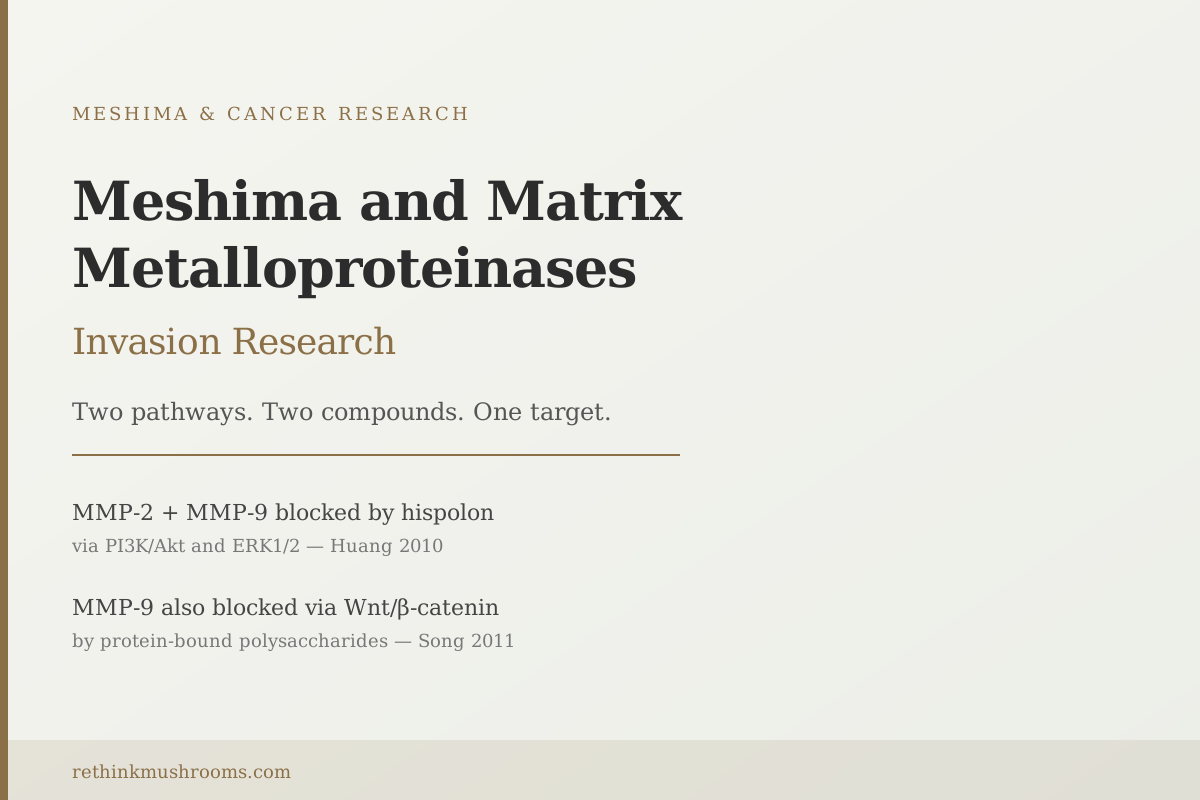
MMP-2 + MMP-9
both gelatinases blocked by hispolon via PI3K/Akt and ERK1/2 (Huang 2010)
Wnt/β-catenin
the second independent pathway through which polysaccharides suppress MMP-9 (Song 2011)
3 cancer types
hepatoma, colon, and thyroid all show MMP inhibition in cell studies
Han et al., International Immunopharmacology, 2006
 View as image ↗
Cancer cells cannot invade surrounding tissue on their own. To move through the protein mesh that holds tissues together, they need molecular tools that dissolve it. Matrix metalloproteinases are those tools: enzymes that cancer cells produce to create physical pathways through what would otherwise be an impassable barrier. Without them, invasion stalls.
Meshima (Phellinus linteus) is a Korean medicinal mushroom that produces compounds studied for their ability to suppress these enzymes. The research identifies two separate mechanisms: a phenolic compound called hispolon that blocks both MMP-2 and MMP-9 through the PI3K/Akt and ERK1/2 signaling pathways,[1] and protein-bound polysaccharides that suppress MMP-9 through Wnt/beta-catenin disruption in an entirely different cell type.[2] The same cancer cell invasion program being targeted through two independent molecular routes.
All of this data comes from cell culture and animal studies. No clinical trials have measured MMP endpoints in humans taking meshima extracts. For a broader overview of meshima's cancer research and what it means in context, see what is meshima mushroom. For research on MMP-9 specifically, a dedicated article covers meshima and MMP-9 inhibition in greater depth.
Key Takeaways
- Matrix metalloproteinases (MMPs) are enzymes cancer cells secrete to dissolve the protein barriers between tissue layers; without them, cancer cells cannot invade or enter the bloodstream
- Hispolon, a phenolic compound from meshima, significantly reduced MMP-2 and MMP-9 expression in human hepatoma cells by suppressing the FAK signaling pathway and its downstream branches: PI3K/Akt/RhoA and ERK1/2 (Huang et al., 2010)
- Protein-bound polysaccharides from meshima inhibit MMP-9 via a completely different route: Wnt/beta-catenin disruption in SW480 colon cancer cells, reducing both MMP-9 activity and cell motility in vitro, and slowing tumor growth in xenograft mice (Song et al., 2011)
- Yu et al. (2022) found that PL polysaccharides reduced MMP expression in thyroid carcinoma cells and modulated epithelial-mesenchymal transition markers, showing MMP inhibition across a third independent cancer type
- Meshima also suppresses urokinase-plasminogen activator (uPA), a related invasion enzyme that works in parallel to MMPs; blocking both creates redundancy in the anti-invasion effect (Sliva et al., 2008)
- All MMP inhibition data for meshima comes from cell assays and animal models; no human trial has measured MMP endpoints after meshima treatment
What MMPs Are: The Enzymes Cancer Uses to Dissolve Its Barriers
To understand why MMP inhibition matters in cancer research, it helps to understand what tissue barriers are.
The body's tissues are separated by thin layers of structural protein. The most important of these, for cancer invasion, is the basement membrane, a dense sheet of type IV collagen and other proteins that underlies every epithelial surface, lines blood and lymphatic vessels, and physically separates tissue compartments from each other. It acts as a barrier. Cancer cells on one side cannot simply push through it. They have to dissolve it.
Matrix metalloproteinases are the enzymes that do the dissolving. They are zinc-dependent proteases, meaning they use a zinc ion at their active site to break peptide bonds in structural proteins. Cancer cells secrete them into the surrounding space, where they degrade the extracellular matrix (ECM), the protein scaffold holding tissue architecture together. As the ECM breaks down, cancer cells move through the gaps.
MMP-2 and MMP-9 are the specific MMPs most commonly elevated in invasive cancers. Both are gelatinases: their primary substrate is denatured collagen and, critically, type IV collagen, making them the two enzymes most directly associated with basement membrane degradation. Elevated MMP-2 and MMP-9 levels in tumor tissue consistently correlate with metastatic potential across multiple cancer types. They are among the most studied drug targets in cancer biology.
Meshima inhibits these two enzymes through at least two independent mechanisms:
- Hispolon blocks MMP-2 and MMP-9 via the PI3K/Akt and ERK1/2 signaling pathways in liver cancer cells (Huang et al., 2010)
- Protein-bound polysaccharides suppress MMP-9 via Wnt/beta-catenin disruption in colon cancer cells (Song et al., 2011)
What makes this notable from a research standpoint: the same molecular target (MMP-9 in particular) is being hit through two independent signaling pathways, by two chemically distinct compounds, in two different cancer cell types.
Hispolon and the Simultaneous Block of MMP-2 and MMP-9
Hispolon is a polyphenol found in Phellinus linteus. In 2010, Huang and colleagues tested it against SK-Hep1 cells, a human hepatocellular carcinoma (liver cancer) line commonly used to study invasion because of its high MMP-2 and MMP-9 expression.[1]
At increasing concentrations of hispolon, expression of MMP-2, MMP-9, and uPA (urokinase-plasminogen activator, a related invasion enzyme) decreased in a dose-dependent way. The cells became less invasive in migration assays.
The study then worked backwards to identify the signaling pathways involved. Focal adhesion kinase (FAK) is a protein that sits at the cell's contact points with the extracellular matrix, sensing attachment and transmitting survival and motility signals inward. When FAK is active, it feeds into two downstream branches: PI3K/Akt/RhoA and ERK1/2. Both branches ultimately increase MMP production.
Hispolon suppressed FAK activity. Both downstream branches quieted: PI3K/Akt, RhoA, and ERK1/2 all showed reduced activity. To confirm that ERK1/2 specifically was driving MMP-2 and MMP-9 expression, the researchers used PD98256, a selective ERK1/2 inhibitor. Applying PD98256 to the cells independently reduced MMP-2 and MMP-9, confirming that ERK1/2 activity was maintaining MMP expression in these cells and that hispolon's effect on MMPs ran through this pathway.
The result is a mechanistically grounded explanation. Hispolon quiets FAK. FAK silencing reduces ERK1/2 and Akt activity. Reduced ERK1/2 and Akt produce less MMP-2 and MMP-9. Less MMP means less ECM degradation, less invasion.
The compound also reduced uPA expression, a protease from a different enzyme family (serine proteases rather than metalloproteinases) that cancer cells use for invasion through a parallel mechanism. The breadth of the effect, hitting two different protease families through the same signaling suppression, is part of what made this study notable.
For more on hispolon and the full range of meshima's bioactive compounds, see Phellinus linteus bioactive compounds: polysaccharides and phenols.
How Polysaccharides Hit MMP-9 Through a Completely Different Route
Hispolon is one compound from meshima. Protein-bound polysaccharides are another, chemically unrelated fraction. Song and colleagues tested them in 2011 against SW480 colon cancer cells.[2]
The signaling pathway involved here is completely different from the FAK/Akt/ERK mechanism that hispolon uses. The Wnt/beta-catenin pathway is a major cell growth and survival signal. In normal cells, beta-catenin is tightly regulated. In many cancers, especially colorectal cancers, Wnt/beta-catenin becomes constitutively active, driving tumor cell proliferation, survival, and invasion. Cyclin D1, a protein that drives cell division, is one of the downstream targets of active beta-catenin. MMP-9 is another.
At concentrations of 125 to 1000 micrograms per milliliter of PL polysaccharide, Song et al. saw: reduced beta-catenin expression, reduced cyclin D1 expression, reduced MMP-9 activity, and reduced cell motility in migration assays. Cell proliferation also slowed significantly.
The study extended to a xenograft mouse model: SW480 cells were implanted in nude mice, and PL polysaccharide treatment significantly slowed tumor growth compared to controls. In the tumor tissue itself, beta-catenin expression was decreased and apoptosis (programmed cell death) was increased. This is important because the in vitro MMP-9 inhibition translated into an in vivo growth-suppression result, not just a cell assay number.
In 2022, Yu and colleagues found the same pattern in a third cancer type: thyroid carcinoma.[3] PL polysaccharide treatment decreased MMP expression and also modulated what are called epithelial-mesenchymal transition (EMT) markers.
EMT is a biological program by which cancer cells shed their original epithelial identity and adopt a more migratory, mesenchymal character. It is one of the molecular switches that transforms a locally contained cancer into an invasive and metastatic one. The key markers are E-cadherin (a protein that keeps epithelial cells anchored to each other, whose loss is associated with invasion) and N-cadherin and Vimentin (proteins that appear when cells shift to a migratory phenotype). PL polysaccharide treatment in thyroid carcinoma cells increased E-cadherin (the anchoring protein) and decreased N-cadherin and Vimentin (the migratory proteins). The cells were being steered back toward an epithelial character.
MMP inhibition and EMT reversal are related but distinct. Blocking MMP-9 slows ECM degradation mechanically. EMT reversal changes what the cancer cell wants to do biologically. The Yu 2022 findings suggest meshima polysaccharides may be doing both.
Beyond MMP: Meshima Also Targets the Invasion Enzyme uPA
Matrix metalloproteinases are not the only enzymes cancer cells use to invade. Urokinase-plasminogen activator, known as uPA, is a serine protease that converts plasminogen to plasmin, a broad-spectrum protease that degrades multiple ECM components and activates latent MMPs. Cancer cells that express high uPA are associated with aggressive invasion and poor prognosis.
Sliva and colleagues tested Phellinus linteus extract against MDA-MB-231 breast cancer cells in 2008, a cell line chosen because it is one of the most invasive in the standard breast cancer model panel.[4] PL treatment significantly reduced uPA secretion. Cell adhesion, migration, and invasion all decreased.
The study also documented suppression of AKT phosphorylation at two sites (Thr308 and Ser473), which are the key activation sites of the AKT protein. Active AKT promotes cell survival, migration, and invasion. In parallel with uPA reduction, the breast cancer cells became less invasive on both measures.
There was an additional finding from the same study: PL inhibited VEGF-mediated capillary formation in an angiogenesis assay (the growth of new blood vessels that tumors need to grow and that can serve as invasion routes). For the broader angiogenesis angle, see how meshima starves tumors: angiogenesis research.
The significance for the MMP story: when cancer cells use uPA to activate latent MMPs in the extracellular environment, blocking uPA secondarily reduces MMP activity. Hispolon blocks MMP-2 and MMP-9 directly at the gene expression level. PL extracts also block uPA at the secretion level, which removes one of the activation signals for MMPs in the tissue environment. The two mechanisms are complementary.
From Cells to Mice: Connecting MMP Research to a Real Metastasis Outcome
Cell culture experiments show that meshima compounds reduce MMP expression, reduce uPA secretion, and reduce invasion in migration assays. But cell assays measure invasion through a thin gel layer in a dish, not through actual tissue. The Han 2006 study moves the evidence into a more meaningful model.[5]
Han and colleagues tested PL acidic polysaccharide in B16F10 melanoma-bearing mice, an animal model used specifically to study metastasis rather than primary tumor growth. B16F10 cells are chosen because they are highly prone to forming lung colonies after injection. At doses of 10, 30, and 100 mg/kg of PL polysaccharide, pulmonary metastasis frequency dropped by 30%, 77%, and 80% respectively.
The mechanism the study identified was not cytotoxic: the polysaccharides were not killing the cancer cells. They were blocking cancer cells from adhering to the extracellular matrix in the target tissue. The ability of cancer cells to attach to the ECM is the step that comes before invasion and colonization: if a traveling cancer cell can't anchor, it can't establish a new tumor. Alongside the adhesion effect, the study documented immunopotentiation: PL polysaccharide increased macrophage nitric oxide (NO) production, activating immune cells that can attack circulating tumor cells.
This dual mechanism (direct adhesion blocking plus immune activation) is consistent with the MMP inhibition data. Less MMP activity means less ECM degradation at the invasion site. Less adhesion means fewer cells successfully seeding new locations. Immune activation adds a third layer of defense against circulating cancer cells.
For the full metastasis research picture, see meshima and cancer metastasis: can it prevent spread?. For the cancer cell migration angle across multiple cancer types, see Phellinus linteus and cancer cell migration inhibition.
What a 2023 Systematic Review Concludes About the Evidence Base
A 2023 systematic review by Qin and colleagues synthesized the extraction, purification, structure, and biological activity of PL polysaccharides across the published literature.[6] It covers anti-tumor, immunomodulatory, and anti-inflammatory functions, confirming that these activities are polysaccharide-dependent and that the structural features of the polysaccharide (branching pattern, molecular weight, degree of sulfonation) influence biological activity.
The review is useful as a synthesis document but also illustrates the state of the field honestly: the evidence base is entirely preclinical. Cell culture and animal models predominate. The molecular mechanisms have been characterized in increasing detail over two decades of laboratory work. What has not happened is the translation into clinical trials with MMP endpoints.
This is important context. MMPs have been the subject of considerable drug development effort, and that history is instructive. Broad-spectrum MMP inhibitor drugs (compounds that block many MMPs at once) failed in multiple clinical trials in the early 2000s, causing musculoskeletal side effects and failing to improve survival despite promising preclinical data. The lesson from that experience was that the timing, selectivity, and cancer stage matter enormously when targeting MMPs. Meshima compounds appear to suppress MMP-2 and MMP-9 rather than blocking all metalloproteinases nonspecifically, which is mechanistically different from the failed broad-spectrum drugs, but whether that selectivity translates to a clinical advantage is unknown.
No clinical trial data exists for meshima and MMP endpoints. The preclinical evidence is consistent across cell types and studies, the molecular mechanisms are well-characterized, and the animal metastasis reduction data provides a meaningful outcome measure. The gap between that and proven clinical benefit remains.
For primary tumor containment research (cell cycle arrest, apoptosis, and chemotherapy synergy), see how meshima may help contain primary tumors.
The Bottom Line
Meshima contains two distinct compound classes that inhibit matrix metalloproteinases (MMPs) through independent signaling pathways: hispolon suppresses MMP-2 and MMP-9 via FAK/PI3K/Akt/RhoA and ERK1/2 in hepatoma cells, while protein-bound polysaccharides suppress MMP-9 via Wnt/beta-catenin disruption in colon cancer cells. A 2022 study in thyroid carcinoma added MMP inhibition alongside EMT marker reversal. Animal data shows 80% lung metastasis reduction at the highest dose tested. All evidence is preclinical; clinical trials measuring MMP endpoints in humans have not been conducted for meshima.
Frequently Asked Questions
Sources & References
- 1RTHM-00344·Huang, Guan-Jhong et al. (2010). “Hispolon suppresses SK-Hep1 human hepatoma cell metastasis by inhibiting matrix metalloproteinase-2/9 and urokinase-plasminogen activator through the PI3K/Akt and ERK signaling pathways..” Journal of agricultural and food chemistry.Study breakdown →PubMed →↩
- 2RTHM-00437·Song, Kyoung-Sub et al. (2011). “Protein-bound polysaccharide from Phellinus linteus inhibits tumor growth, invasion, and angiogenesis and alters Wnt/β-catenin in SW480 human colon cancer cells..” BMC cancer.Study breakdown →PubMed →↩
- 3RTHM-01643·Yu, Kun et al. (2022). “Systematic evaluation of the anti-tumor effect of Phellinus linteus polysaccharide in thyroid carcinoma in vitro..” Molecular biology reports.Study breakdown →PubMed →↩
- 4RTHM-00271·Sliva, D et al. (2008). “Phellinus linteus Extract in Breast-Cancer Cell Models: What This 2008 Study Actually Found.” British journal of cancer.Study breakdown →PubMed →↩
- 5RTHM-00185·Han, Sang-Bae et al. (2006). “Acidic polysaccharide from Phellinus linteus inhibits melanoma cell metastasis by blocking cell adhesion and invasion..” International immunopharmacology.Study breakdown →PubMed →↩
- 6RTHM-01770·Qin, Dandan et al. (2023). “Polysaccharides from Phellinus linteus: A systematic review of their extractions, purifications, structures and functions..” International journal of biological macromolecules.Study breakdown →PubMed →↩