Mushrooms & Brain Health
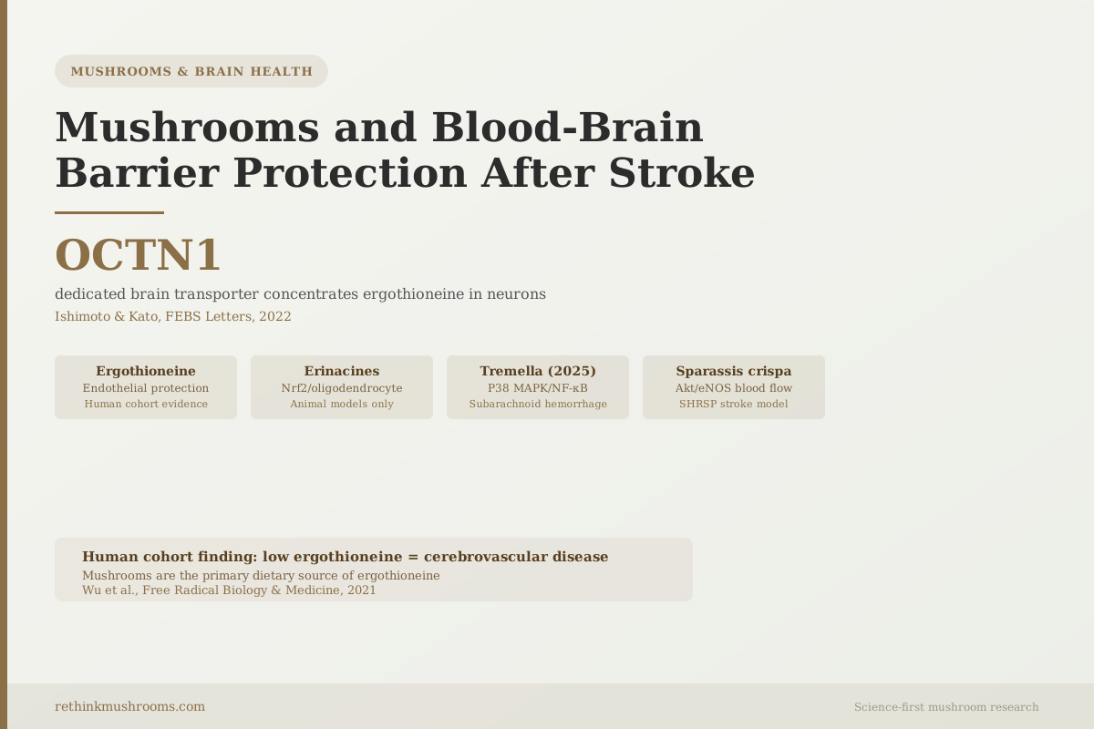
OCTN1: a dedicated brain transporter concentrates ergothioneine in neurons
Ergothioneine, found almost exclusively in mushrooms, has a dedicated high-affinity transporter (OCTN1/SLC22A4) that carries it into brain parenchymal cells including neurons, neural stem cells, and microglia. Low plasma ergothioneine is associated with cerebrovascular disease in human patients.
Ishimoto & Kato, FEBS Letters, 2022; Wu et al., Free Radical Biology & Medicine, 2021
496 participants
in a cross-sectional study, plasma ergothioneine was lowest in dementia (p < 0.001) and low levels tracked with white matter hyperintensities and brain atrophy — the only human data linking this mushroom compound to cerebrovascular disease (Wu et al., Free Radical Biology & Medicine, 2021)
Sparassis crispa
delayed stroke incidence and death in stroke-prone hypertensive rats by activating Akt-dependent eNOS and increasing cerebral blood flow (Yoshitomi et al., Journal of Natural Medicines, 2011)
2025: Tremella fuciformis
polysaccharides reduced early brain injury in subarachnoid hemorrhage by inhibiting the KDR-P38 MAPK/NF-κB inflammatory cascade (Wan et al., Molecular Neurobiology, 2025)
Ishimoto & Kato, FEBS Letters, 2022; Wu et al., Free Radical Biology & Medicine, 2021
 View as image ↗
When a stroke occurs, the visible damage, the infarct and dead tissue, is not the end of the injury. What follows is a cascade of secondary damage driven partly by the breakdown of the blood-brain barrier. The barrier's collapse lets inflammatory cells and fluid enter the brain from the bloodstream, worsening edema and killing additional neurons that survived the initial ischemia. Protecting the barrier, or limiting the damage from its disruption, is one of the least-discussed angles in both conventional and natural medicine for stroke recovery. This article covers what the research shows for mushroom compounds in that specific context. For the full picture of mushrooms and brain health, see the complete guide to mushrooms and cognitive aging.
Key Takeaways
- Ergothioneine, found almost exclusively in mushrooms, has a dedicated brain transporter (OCTN1/SLC22A4) that carries it into brain parenchymal cells including neurons, neural stem cells, and microglia; knockout mice lacking OCTN1 have no brain ergothioneine at all (Ishimoto & Kato, FEBS Letters, 2022)
- Low plasma ergothioneine was significantly associated with both neurodegeneration and cerebrovascular disease in a cross-sectional study of 496 people, the closest thing to direct human evidence linking mushroom compound status to stroke-related brain injury — though its design cannot establish which came first (Wu et al., Free Radical Biology & Medicine, 2021)
- Ergothioneine protected hCMEC/D3 human brain endothelial cells — the cell type that forms the physical structure of the blood-brain barrier — from 7-ketocholesterol-induced viability loss, apoptosis and necrosis, though in isolated culture rather than an intact barrier (Koh et al., Neuromolecular Medicine, 2021)
- Erinacines from lion's mane mycelium protected the brain from traumatic injury via Nrf2-dependent pathways in animal models (Lee et al., Antioxidants, 2024), but no human pharmacokinetic data confirms these compounds reach neurons at therapeutic concentrations in people
- Tremella fuciformis polysaccharides reduced cerebral edema and improved neurological function 24 hours after experimental subarachnoid hemorrhage in mice, the most recent study in this literature — though a hemorrhagic model addresses a different stroke subtype than ischemic stroke (Wan et al., Molecular Neurobiology, 2025)
- Sparassis crispa delayed stroke incidence and death in stroke-prone hypertensive rats fed 1.5% of the mushroom in their diet, raising cerebral phospho-eNOS and phospho-Akt and increasing blood flow (Yoshitomi et al., 2011) — the only published stroke study for this species, and a prevention study rather than a recovery one
- Not one of the fourteen references cited here measured tight junction proteins or barrier permeability directly. The closest measured endpoints are cerebral edema in two rodent studies (Pang et al., 2020; Wan et al., 2025) and endothelial cell survival in a dish (Koh et al., 2021)
- No human clinical trial has tested any mushroom compound for blood-brain barrier protection, edema, or neurological outcome after stroke. Stroke is a medical emergency and nothing here is a treatment or a reason to delay emergency care
Read First
CriticalStroke Is a Medical Emergency — Nothing Here Is a Treatment
Concern
Stroke care is time-critical: the window for clot-dissolving therapy in ischemic stroke is measured in hours, and delay costs surviving brain tissue. Every mechanistic finding described in this article comes from cell cultures or animal models, and the single human study is an observational snapshot in dementia patients, not stroke patients. Not one of these compounds has been given to a human being and tested for blood-brain barrier protection after stroke.
What the research says
Treat suspected stroke as an emergency and call emergency services immediately — sudden face drooping, arm weakness, or speech difficulty are the standard warning signs. No mushroom, extract, or supplement substitutes for emergency care, and none should delay it by a single minute.
Particularly relevant for: Anyone who has had a stroke, is at elevated risk of one, or is currently on post-stroke medication
What to do
If you have had a stroke and want to add mushrooms to your diet or take a supplement, raise it with the physician managing your stroke care first. Post-stroke patients are typically on antiplatelet or anticoagulant regimens, and no study cited here tested a mushroom compound alongside those medications — the interaction profile is simply unknown, not established as safe.
Evidence base reviewed in this article: fourteen references — ten primary studies and four reviews. Wu et al. (2021) is the sole primary study in humans and is observational; every other primary study is preclinical (cell culture or animal model). This article is educational and is not medical advice.
What the Blood-Brain Barrier Is and Why Stroke Damages It
The blood-brain barrier is a selective physical boundary between the bloodstream and the brain tissue. It is composed of specialized endothelial cells, the cells that line blood vessels, held together by tight junction proteins that create an unusually impermeable seal. Unlike blood vessels in most of the body, the brain's vessels do not allow large molecules, most pathogens, or immune cells to pass freely from blood to brain tissue. This selectivity is what protects the brain's delicate chemical environment.
During a stroke, this barrier breaks down. In ischemic stroke, reduced blood flow triggers a cascade that begins with energy failure in endothelial cells. Within 4 to 6 hours, the tight junction proteins between endothelial cells are degraded by enzymes called matrix metalloproteinases. Once the tight junctions fail, the barrier becomes permeable. Fluid, inflammatory cells, and cytokines from the bloodstream enter the brain. This creates edema (brain swelling), amplifies neuroinflammation, and extends the zone of cell death well beyond the initial ischemic core.
This secondary injury from BBB breakdown accounts for a substantial portion of post-stroke disability. Interventions that protect endothelial cell integrity, reduce tight junction degradation, or limit the inflammatory infiltration that follows disruption are theoretically among the most impactful approaches to stroke outcome.
Several mushroom compounds target these endothelial cell biology and neuroinflammation pathways. The most specific and clinically grounded evidence involves ergothioneine.
Cascade · Intervention Point · Evidence
Where Each Mushroom Compound Acts — and the Step Nobody Measured
Blood-brain barrier failure after stroke is a sequence, not an event. Mapping every cited compound onto the step it was actually shown to affect reveals the shape of this literature: well populated at perfusion and at inflammation, and completely empty at the tight-junction step that is the definition of barrier breakdown.
0.Before the stroke — perfusion and vessel tone
Chronic hypertension and endothelial dysfunction raise stroke risk and worsen outcome when one occurs.
- Sparassis crispaRef 9
Lowered blood pressure, raised cerebral blood flow, increased phospho-eNOS and phospho-Akt in cortex; delayed stroke onset and death in SHRSP rats
1.Ischemia → endothelial energy failure
Blood flow stops. The endothelial cells lining brain vessels lose ATP and begin to fail. Reperfusion then floods the tissue with reactive oxygen species.
- ErgothioneineRef 5
Protected hCMEC/D3 human brain endothelial cells from 7-ketocholesterol-induced viability loss, apoptosis and necrosis — in culture, not in a stroke
- ErinacinesRef 6
Nrf2-dependent antioxidant activation in rat mild traumatic brain injury and primary glial cells — counters reperfusion oxidative stress by mechanism, tested in a different injury
2.Matrix metalloproteinases degrade tight junctions
Within roughly 4–6 hours, MMPs chew through the tight junction proteins that seal the barrier. This is the step that defines barrier failure.
Evidence gapNo cited study measured tight junction proteins or barrier permeability directly. This is the mechanistic centre of the topic and the emptiest rung in the evidence.
3.Barrier leaks → edema and immune infiltration
Fluid, plasma proteins and circulating immune cells cross into brain tissue. The brain swells.
- Pachymic acidRef 11
Reduced brain water content and infarct volume, raised cerebral blood flow after rat ischemia-reperfusion, via PI3K/Akt — brain water content is a direct edema measure
- Tremella polysaccharidesRef 12
Reduced cerebral edema and improved neurological function 24 h after mouse subarachnoid hemorrhage — a hemorrhagic, not ischemic, model
4.Microglial activation and neuroinflammation
Resident microglia switch to a pro-inflammatory state, releasing cytokines that sustain the damage and keep the barrier open.
- Ganoderma polysaccharidesRef 10
Attenuated microglia-mediated neuroinflammation, modulated microglial phagocytosis and behavioural response
- Tremella polysaccharidesRef 12
Reversed microglial activation and enhanced phagocytic capacity via KDR-mediated P38 MAPK/NF-κB inhibition
5.Secondary neuron death and demyelination
Neurons that survived the initial ischemia die. Oligodendrocytes are lost and myelin degrades, impairing signal conduction and recovery.
- Erinacine SRef 7
Protected oligodendrocytes and alleviated mood abnormalities in cuprizone-exposed rodents — a demyelination model, not stroke
- Hericium myceliumRef 8
Promoted oligodendrocyte maturation with increased myelin basic protein in OPC culture and ex vivo cerebellar slices
Every entry is preclinical. Three of the seven compound placements come from models that are not stroke at all — mild traumatic brain injury, cuprizone demyelination, and cell culture — and one comes from hemorrhagic rather than ischemic stroke.
Source: Yoshitomi et al. (2011); Koh et al. (2021); Lee et al. (2024); Pang et al. (2020); Wan et al. (2025); Cai et al. (2017); Fu et al. (2024); Huang et al. (2021). Cascade sequence as described in the article text.
The map above is worth sitting with before reading the compound-by-compound evidence, because it frames what follows. The research is real, but it is distributed unevenly across the cascade — and the gap sits exactly where the topic's name points.
Ergothioneine: The Mushroom Compound With a Dedicated Brain Transporter
Most dietary antioxidants get into the brain by passive diffusion or by chance, if they are small and lipophilic enough to pass through the lipid-rich BBB endothelial cell membranes. Ergothioneine is different. The brain actively collects it.
Ishimoto and Kato described this in detail in a 2022 FEBS Letters review: ergothioneine is extremely hydrophilic, the kind of molecule that should struggle to enter the brain at all, yet it is absorbed from the gut and distributed to the brain because the ergothioneine-specific transporter OCTN1 (also designated SLC22A4) carries it into brain parenchymal cells — neurons, neural stem cells, and microglia among them.[2] The cleanest demonstration is genetic: mice with the Octn1 gene knocked out have no ergothioneine in the brain whatsoever. Remove the transporter and the compound simply does not arrive.
The evolutionary significance of a dedicated high-affinity brain transporter is generally interpreted as evidence that the compound serves important biological functions in that tissue. Cells do not maintain energy-dependent transporters for compounds that are incidental.
What happens when ergothioneine is low? Wu and colleagues provided the most direct human evidence in 2021, measuring plasma ergothioneine by mass spectrometry across 496 participants: 88 with no cognitive impairment, 201 with cognitive impairment but no dementia, and 207 with dementia (160 Alzheimer's disease, 47 vascular dementia).[1] Plasma ergothioneine was lowest in the dementia group (p < 0.001 versus both other groups), with intermediate levels in the cognitive-impairment group (p < 0.001 versus unimpaired) — a stepwise decline tracking disease severity rather than a simple present-or-absent difference.
The cerebrovascular specifics matter for this article. In multivariate models adjusted for demographic and vascular risk factors, lower ergothioneine was significantly associated with dementia both with and without cerebrovascular disease, while the association with cognitive impairment short of dementia held only in the presence of cerebrovascular disease. Lower ergothioneine also tracked with white matter hyperintensities and with brain atrophy markers — reduced global cortical thickness and smaller hippocampal volumes. White matter hyperintensities are a recognized imaging signature of small-vessel cerebrovascular damage, which makes this the closest the literature comes to connecting a mushroom compound to the vascular integrity of a living human brain.
The design caveat is real and should not be glossed. This is a cross-sectional study: every measurement was taken at a single point in time, so it cannot establish whether low ergothioneine contributed to the brain damage or resulted from it. Illness, appetite loss, and dietary change all plausibly lower ergothioneine intake in people who already have dementia, and the study cannot rule that direction out. What it establishes is a robust, dose-graded association — not a causal or therapeutic relationship. Given that mushrooms are by far the primary dietary source of ergothioneine, the finding is still the most practically relevant human data point available.
Wu et al. (2021) · Free Radical Biology & Medicine
- Population
- 496 participants: 88 cognitively unimpaired, 201 with cognitive impairment but no dementia, 207 with dementia (160 Alzheimer's, 47 vascular dementia)
- Intervention
- No intervention — plasma ergothioneine and its metabolite l-hercynine measured once by LC-MS/MS, alongside neuropsychological testing and neuroimaging for cerebrovascular disease and brain atrophy
- Finding
- Plasma ergothioneine was lowest in dementia (p < 0.001 vs. both other groups) and intermediate in cognitive impairment (p < 0.001 vs. unimpaired). After adjusting for demographic and vascular risk factors, low ergothioneine was significantly associated with dementia with and without cerebrovascular disease, and separately with white matter hyperintensities, reduced cortical thickness, and smaller hippocampal volumes.
narrow-scope Cross-sectional, so it cannot establish direction. Dementia itself can reduce appetite and dietary intake, which would lower ergothioneine as a consequence rather than a cause. This is the only human study in this entire article, and it measures association in dementia patients — not blood-brain barrier protection, and not stroke.
Wu, Cheah, Chong et al. (2021). Free Radical Biology & Medicine, 177, 201-211. Reference 1.
Koh and colleagues provided the most mechanistically specific BBB evidence in 2021, testing ergothioneine against 7-ketocholesterol-induced injury in hCMEC/D3 cells, an immortalized human brain endothelial cell line.[5] 7-ketocholesterol is an oxidized cholesterol derivative that accumulates in patients with cardiovascular disease and diabetes and is thought to drive pathological inflammation; in these cells it caused a dose-dependent loss of viability with increased apoptosis and necrosis. Ergothioneine protected the endothelial cells from that injury. The significance is the cell type: hCMEC/D3 is a human brain endothelial line, meaning the cells that form the physical structure of the blood-brain barrier were the ones protected. The limitation is equally specific — this is a cell line in a dish, exposed to a single oxidized cholesterol species, not a barrier in a living brain during a stroke.
Paul's 2022 review framed ergothioneine's role as a "stress vitamin" with three relevant properties for vascular and cerebrovascular health: antiaging effects on vascular tissue, direct endothelial protection, and neuroprotective effects in CNS tissues.[4] Nakamichi and colleagues' 2022 review described the same transport architecture from a different angle — OCTN1 expressed in brain parenchymal cells including neurons, blood ergothioneine permeating the blood-brain barrier into brain tissue — and reviewed ergothioneine's neuroprotective roles specifically in CNS diseases, including protection against the oxidative stress that drives post-stroke secondary injury.[3] They also note that ergothioneine is unusually stable in the body, remaining for a long period after ingestion.
The practical implication is narrower than it first appears. Humans cannot synthesise ergothioneine and take it up almost entirely from the diet, with mushrooms as the dominant source.[5] Among mushrooms, oyster and king oyster rank high and porcini higher still — the species comparison is covered in detail in the dedicated ergothioneine research below. But the honest framing of what eating them buys you is this: a plausible mechanism plus an observational association, not demonstrated cerebrovascular protection. Eating mushrooms is a reasonable dietary habit on this evidence. It is not an intervention with a proven vascular endpoint.
For the detailed ergothioneine brain research, see ergothioneine and brain health: neuroprotective research.
Erinacines and the BBB: Real Animal Evidence, Unconfirmed in Humans
Erinacines are diterpenoid compounds produced by lion's mane mycelium, distinct from the hericenones found in the fruiting body. Erinacines are the compounds carrying the barrier-crossing evidence: a 2025 narrative review characterizes erinacine-A-enriched Hericium erinaceus as having the highest bioactive potency among the mushroom's extracted compounds and as showing comparative ease of transport across biological barriers.[14] That is a review-level characterization rather than a direct pharmacokinetic measurement, and the distinction matters for everything that follows in this section.
Lee and colleagues demonstrated in 2024 that novel erinacines from Hericium erinaceus mycelium protected the brain in a mild traumatic brain injury model in vivo and protected primary glial cells from neuroinflammation via Nrf2-dependent antioxidant pathways.[6] Nrf2 activation upregulates the cell's antioxidant enzyme machinery, directly countering the reactive oxygen species produced when ischemic tissue reperfuses. This mechanism is relevant to stroke because ischemia-reperfusion injury, where blood flow resumes after a blockage and generates oxidative stress, is a major driver of secondary damage.
Erinacine S protects the oligodendrocytes, the cells responsible for producing the myelin sheath that insulates axons. Fu and colleagues documented in 2024 that erinacine S protected oligodendrocytes from demyelination in a cuprizone model.[7] Huang and colleagues showed in 2021 that Hericium erinaceus mycelium and its small compounds promoted oligodendrocyte maturation and increased expression of myelin basic protein.[8] Myelin integrity is part of what keeps axons functional; post-stroke demyelination is one component of the structural damage that impairs recovery.
The caveat matters and is worth stating precisely: erinacines have been shown to cross the BBB in preclinical animal studies. No published human pharmacokinetic data confirms that erinacines reach neurons at therapeutically relevant concentrations in people after oral supplementation. The gap between animal BBB penetration data and human brain tissue concentrations is unknown. Claims that lion's mane "crosses the blood-brain barrier" in humans are extrapolated from animal data, not confirmed by human measurement.
For the dedicated explainer on the BBB-crossing evidence for erinacines, see erinacines: the compounds that cross the blood-brain barrier.
Additional Compounds: Ganoderma, Poria, Tremella, and Sparassis
Several other mushroom species and compounds have preclinical data relevant to post-stroke BBB biology.
Ganoderma lucidum (reishi) polysaccharides attenuated microglia-mediated neuroinflammation in a 2017 study by Cai and colleagues, also modulating microglial phagocytosis and improving behavioral responses in animal models.[10] After stroke, microglia are the main drivers of the neuroinflammatory cascade that follows BBB breakdown. Compounds that switch microglia from a pro-inflammatory activation state to a more regulatory state reduce secondary injury. The Ganoderma polysaccharide data is from animal models, but the mechanism targets the immediate post-stroke biology directly.
Pachymic acid is a lanostane triterpenoid from Poria cocos (Fu Ling, a fungal ingredient used extensively in traditional Chinese medicine). Pang and colleagues demonstrated in 2020 that pachymic acid significantly reduced cerebral ischemia-reperfusion injury in rats by activating the PI3K/Akt signaling pathway, a pro-survival cascade that promotes neuron survival after energy stress.[11] The endpoints are worth listing because they are the most barrier-relevant in this article: pachymic acid increased cerebral blood flow, reduced infarct volume, reduced neuronal apoptosis on TUNEL staining, and reduced brain water content. That last measure is the closest any cited study comes to quantifying a leaking barrier — brain water content is how edema is measured, and edema is what happens when the barrier fails. Blocking PI3K with LY294002 abolished the effect, which is the kind of control that makes the proposed mechanism credible rather than merely correlated. It remains a rat study.
The most recent mushroom BBB data comes from Wan and colleagues' 2025 study in Molecular Neurobiology, testing Tremella fuciformis polysaccharides in an experimental subarachnoid hemorrhage model in mice.[12] Subarachnoid hemorrhage is a severe form of hemorrhagic stroke associated with very high rates of early brain injury and death. Twenty-four hours after the hemorrhage, treated mice showed improved neurological function and reduced cerebral edema — edema being the direct downstream consequence of a leaking barrier. Mechanistically, the polysaccharides reversed microglial activation, enhanced microglial phagocytic capacity, and reduced neuronal apoptosis, working through the KDR receptor-mediated P38 MAPK/NF-κB cascade identified by network pharmacology and then confirmed by flow cytometry, Western blotting, and immunofluorescence. P38 MAPK is one of the kinase pathways that drives tight junction protein degradation after BBB disruption. Worth noting: this is a hemorrhagic model, so it speaks to a different stroke subtype than the ischemic injury most of this article addresses.
Sparassis crispa, a cauliflower-like fungal species, produced one of the most striking results in this literature. Yoshitomi and colleagues demonstrated in 2011 that feeding stroke-prone spontaneously hypertensive rats a diet containing 1.5% Sparassis crispa delayed the incidence of stroke and death, significantly decreased blood pressure, and increased blood flow.[9] After four weeks of feeding, the cerebral cortex of treated rats showed increased phosphorylated eNOS (Ser1177) and phosphorylated Akt (Ser473), along with higher nitrate/nitrite concentrations in cerebral tissue — direct biochemical evidence that the Akt/eNOS pathway was activated and producing more nitric oxide. eNOS is the enzyme that produces nitric oxide in endothelial cells; nitric oxide relaxes blood vessels, improves perfusion, and has direct endothelial-protective effects. This is the only published stroke study for Sparassis crispa. Its strength is that the SHRSP model is purpose-built for stroke research and the endpoint was actual stroke incidence, not a surrogate marker; its limitation is that the intervention was preventive, started in young rats before any stroke occurred, which is a fundamentally different question from protecting a barrier after a stroke has already happened.
Yamashina and colleagues demonstrated in 2022 that fruiting bodies of medicinal mushrooms suppressed demyelination in a cuprizone-induced model, feeding mice a diet containing 5% mushroom for five weeks.[13] Four species — Pleurotus eryngii, Ganoderma lucidum, Flammulina velutipes, and Hericium erinaceus — effectively suppressed demyelination, and three of them (P. eryngii, G. lucidum, F. velutipes) significantly improved cuprizone-induced motor dysfunction. This is a multiple sclerosis model rather than a stroke model, so the relevance to post-stroke recovery is indirect: it demonstrates that these species can protect myelin under chemical insult, not that they repair myelin damaged by ischemia.
For the full multi-compound and multi-species preclinical evidence for cerebral ischemia, see mushroom compounds and cerebral ischemia: preclinical evidence. For the inflammation angle specifically, see can mushrooms help after a stroke: anti-inflammatory brain effects.
What the Evidence Does and Doesn't Show
- Cell studiescompleted
Ergothioneine protected human brain endothelial cells (hCMEC/D3) from 7-ketocholesterol injury (Koh 2021). Hericium mycelium compounds promoted oligodendrocyte maturation in OPC culture (Huang 2021).
- Animal studiescurrent
Seven of the ten primary studies cited here. Sparassis crispa delayed stroke in SHRSP rats (Yoshitomi 2011); pachymic acid reduced infarct volume and brain water content in rat ischemia-reperfusion (Pang 2020); Tremella polysaccharides reduced edema in mouse subarachnoid hemorrhage (Wan 2025); erinacines protected via Nrf2 in rat mild TBI (Lee 2024); four species suppressed demyelination in cuprizone mice (Yamashina 2022).
- Human observationalcompleted
One study, and it is not about stroke: Wu 2021 linked low plasma ergothioneine to cerebrovascular disease markers in 496 dementia-clinic participants, cross-sectionally.
- Small human trialsmissing
No trial has given a mushroom compound to a stroke patient and measured blood-brain barrier integrity, edema, or neurological outcome.
- Large randomized trialsmissing
None registered or published for this indication.
- Meta-analysismissing
Not possible — there are no human trials to pool.
The mechanistic and animal layers are genuinely populated, and in the case of ergothioneine the biology is unusually well characterized down to a named transporter. But the ladder has a hard break above the animal rung. The single human study is observational, cross-sectional, and measured dementia rather than stroke. Anyone claiming mushrooms protect the blood-brain barrier after a human stroke is describing a rung that does not exist.
Fourteen references cited in this article — ten primary studies (Koh 2021; Huang 2021; Yoshitomi 2011; Pang 2020; Wan 2025; Lee 2024; Fu 2024; Cai 2017; Yamashina 2022; Wu 2021) and four reviews (Ishimoto & Kato 2022; Nakamichi 2022; Paul 2022; Cornford 2025).
With one observational exception, this body of research is entirely preclinical. No human clinical trials have tested any mushroom compound specifically for blood-brain barrier protection after stroke. The exception, Wu 2021, is a cross-sectional study that establishes a human association between low plasma ergothioneine and cerebrovascular disease markers — and a single-timepoint association is not a causal, temporal, or therapeutic relationship. It also studied dementia-clinic patients, not stroke patients.
The erinacine and other lion's mane data comes from animal models of different brain injuries (TBI, demyelination, ischemia) rather than BBB-specific stroke endpoints. The Tremella, Sparassis, Ganoderma, and Poria data each comes from a single animal study per compound.
There is a sharper way to state the limitation, and it comes from asking a simple question of each study: what did it actually measure?
Claim · Endpoint · Distance
Ten Studies, Zero Direct Measurements of the Barrier
“Blood-brain barrier protection” is a specific claim with a specific test: did the tight junctions hold, and did the barrier stay sealed? Sorting every cited study by what it actually measured shows how far the literature sits from that test.
No cited study measured tight junction proteins (claudin, occludin, ZO-1) or barrier permeability. The endpoint that would justify the phrase “barrier protection” has not been tested for any of these compounds in stroke.
- Pang et al., 2020Edema — physiological proxy for leakRef 11
Pachymic acid (Poria cocos)
Rat cerebral ischemia-reperfusion
Measured: Brain water content, infarct volume, cerebral blood flow, neurological scores, neuronal apoptosis (TUNEL), PI3K/Akt proteins
- Wan et al., 2025Edema — physiological proxy for leakRef 12
Tremella fuciformis polysaccharides
Mouse subarachnoid hemorrhage
Measured: Cerebral edema, neurological function at 24 h, microglial activation and phagocytosis, neuronal apoptosis, KDR / P38 MAPK / NF-κB
- Koh et al., 2021Barrier cell survival, in vitroRef 5
Ergothioneine
hCMEC/D3 human brain endothelial cell line
Measured: Cell viability, apoptosis and necrosis under 7-ketocholesterol challenge — the barrier-forming cell type, but isolated in culture
- Yoshitomi et al., 2011Upstream — perfusion and vessel toneRef 9
Sparassis crispa (1.5% of diet)
Stroke-prone spontaneously hypertensive rats
Measured: Stroke incidence and survival, blood pressure, blood flow, cerebral nitrate/nitrite, phospho-eNOS (Ser1177), phospho-Akt (Ser473)
- Cai et al., 2017Downstream — after the barrier failedRef 10
Ganoderma lucidum polysaccharides
Microglia, neuroinflammation model
Measured: Microglia-mediated neuroinflammation, microglial phagocytosis, behavioural response
- Lee et al., 2024Downstream — after the barrier failedRef 6
Erinacines / H. erinaceus mycelium
Rat mild traumatic brain injury + primary glial cells
Measured: Oxidative stress modulation, Nrf2-dependent pathways, neurological improvement
- Fu et al., 2024Downstream — after the barrier failedRef 7
Erinacine S
Cuprizone-exposed rodents
Measured: Oligodendrocyte protection, myelin, mood abnormalities
- Huang et al., 2021Downstream — after the barrier failedRef 8
H. erinaceus mycelium compounds
OPC culture, ex vivo cerebellar slices
Measured: Oligodendrocyte maturation, myelin basic protein expression
- Yamashina et al., 2022Downstream — after the barrier failedRef 13
Four species, 5% of diet, 5 weeks
Cuprizone-induced MS model mice
Measured: Demyelination (Black-Gold II), rotarod motor function, body weight
- Wu et al., 2021Downstream — after the barrier failedRef 1
Ergothioneine (dietary status)
Human, cross-sectional, n = 496
Measured: Plasma ergothioneine vs. white matter hyperintensities, cortical thickness, hippocampal volume — imaging markers of vascular damage, measured once
Reading down the list is reading outward from the claim. Two rodent studies measured edema, which a failing barrier causes. One measured survival of barrier cells in a dish. The rest measured events upstream or downstream of the barrier entirely.
Source: Pang et al. (2020); Wan et al. (2025); Koh et al. (2021); Yoshitomi et al. (2011); Cai et al. (2017); Lee et al. (2024); Fu et al. (2024); Huang et al. (2021); Yamashina et al. (2022); Wu et al. (2021). Endpoints as reported in each published abstract.
What the evidence does show is that multiple mushroom compounds hit mechanistic targets that are directly relevant to post-stroke BBB biology: tight junction protection, endothelial cell survival, microglial modulation, Nrf2-mediated antioxidant defense, and oligodendrocyte preservation. The convergence across species and compound classes is unlikely to be coincidental; it reflects a real biological overlap between mushroom bioactive compounds and the cellular machinery involved in BBB protection.
The most practically grounded finding remains ergothioneine. Dietary intake through oyster mushrooms, king oyster mushrooms, and porcini mushrooms is a low-risk way to maintain the plasma ergothioneine levels that Wu 2021 found associated with fewer cerebrovascular and atrophy markers. Framed honestly, that is a reason to eat mushrooms as part of a normal diet — the same conclusion the evidence would support for most whole foods with favorable observational data. It is not a reason to take a supplement expecting stroke protection, and it is not a substitute for controlling the risk factors with actual causal evidence behind them: blood pressure, atrial fibrillation, smoking, diabetes, and cholesterol.
The Bottom Line
Stroke breaks down the blood-brain barrier within hours, triggering secondary injury that can equal or exceed the initial ischemic damage. The most grounded human evidence for a mushroom-BBB connection involves ergothioneine: a cross-sectional study of 496 people linked low plasma ergothioneine to cerebrovascular disease markers, white matter hyperintensities, and brain atrophy, and the compound has a dedicated transporter (OCTN1/SLC22A4) that carries it into brain parenchymal cells — knockout mice lacking it have no brain ergothioneine at all. Ergothioneine also protected human brain endothelial cells from oxidized cholesterol injury in culture. Additional preclinical evidence spans Tremella fuciformis polysaccharides reducing edema in mouse subarachnoid hemorrhage (2025), pachymic acid reducing brain water content in rat ischemia-reperfusion, Sparassis crispa activating Akt/eNOS in stroke-prone rats, Ganoderma polysaccharides suppressing microglial neuroinflammation, and erinacines protecting oligodendrocytes via Nrf2 pathways. The limitation worth stating plainly: no cited study measured tight junction integrity or barrier permeability directly, and no human clinical trial has tested any of these compounds for BBB protection after stroke. Stroke is a medical emergency; none of this is a treatment.
Frequently Asked Questions
Sources & References
- 1RTHM-01456·Wu, Liu-Yun et al. (2021). “Low plasma ergothioneine levels are associated with neurodegeneration and cerebrovascular disease in dementia..” Free radical biology & medicine.Study breakdown →PubMed →↩
- 2RTHM-01544·Ishimoto, Takahiro et al. (2022). “Ergothioneine in the brain..” FEBS letters.Study breakdown →PubMed →↩
- 3RTHM-01589·Nakamichi, Noritaka et al. (2022). “Ergothioneine and central nervous system diseases..” Neurochemical research.Study breakdown →PubMed →↩
- 4RTHM-01594·Paul, Bindu D (2022). “Ergothioneine: A Stress Vitamin with Antiaging, Vascular, and Neuroprotective Roles?.” Antioxidants & redox signaling.Study breakdown →PubMed →↩
- 5RTHM-01373·Koh, Sally Shuxian et al. (2021). “Effect of Ergothioneine on 7-Ketocholesterol-Induced Endothelial Injury..” Neuromolecular medicine.Study breakdown →PubMed →↩
- 6RTHM-01924·Lee, Kam-Fai et al. (2024). “The Cerebral Protective Effect of Novel Erinacines from Hericium erinaceus Mycelium on In Vivo Mild Traumatic Brain Injury Animal Model and Primary Mixed Glial Cells via Nrf2-Dependent Pathways..” Antioxidants (Basel.Study breakdown →PubMed →↩
- 7RTHM-00878·Fu, Jing-Ting et al. (2024). “Erinacine S, a small active component derived from Hericium erinaceus, protects oligodendrocytes and alleviates mood abnormalities in cuprizone-exposed rodents..” Biomedicine & pharmacotherapy = Biomedecine & pharmacotherapie.Study breakdown →PubMed →↩
- 8RTHM-00692·Huang, Hui-Ting et al. (2021). “Hericium erinaceus mycelium and its small bioactive compounds promote oligodendrocyte maturation with an increase in myelin basic protein..” Scientific reports.Study breakdown →PubMed →↩
- 9RTHM-00446·Yoshitomi, Hisae et al. (2011). “Beneficial effect of Sparassis crispa on stroke through activation of Akt/eNOS pathway in brain of SHRSP..” Journal of natural medicines.Study breakdown →PubMed →↩
- 10RTHM-00462·Cai, Qing et al. (2017). “Polysaccharides from Ganoderma lucidum attenuate microglia-mediated neuroinflammation and modulate microglial phagocytosis and behavioural response..” Journal of neuroinflammation.Study breakdown →PubMed →↩
- 11RTHM-01273·Pang, Yingqiao et al. (2020). “Pachymic acid protects against cerebral ischemia/reperfusion injury by the PI3K/Akt signaling pathway..” Metabolic brain disease.Study breakdown →PubMed →↩
- 12RTHM-02201·Wan, Yu-Hui et al. (2025). “Tremella fuciformis Polysaccharides Alleviate Early Brain Injury in Experimental Subarachnoid Hemorrhage by Inhibiting the KDR-Mediated P38 MAPK/NF-κB Pathway..” Molecular neurobiology.Study breakdown →PubMed →↩
- 13RTHM-01637·Yamashina, Kota et al. (2022). “Suppressive Effect of Fruiting Bodies of Medicinal Mushrooms on Demyelination and Motor Dysfunction in a Cuprizone-Induced Multiple Sclerosis Mouse Model..” International journal of medicinal mushrooms.Study breakdown →PubMed →↩
- 14RTHM-00922·Cornford, Nisha et al. (2025). “Hericium erinaceus: A possible future therapeutic treatment for the prevention and delayed progression of Alzheimer's disease? - A narrative review..” Nutrition research reviews.Study breakdown →PubMed →↩