Lion's Mane & Neuroplasticity
16 weeks to measurable gain
Mori et al. (2009) found lion's mane at 3g/day produced significantly higher cognitive scores in MCI patients versus placebo at weeks 8, 12, and 16. Scores declined after stopping supplementation, consistent with a neurogenesis-driven effect that requires ongoing support.
Mori et al., Phytotherapy Research, 2009
Dentate gyrus
the hippocampal region where lion's mane promotes new neuron growth in adult animals
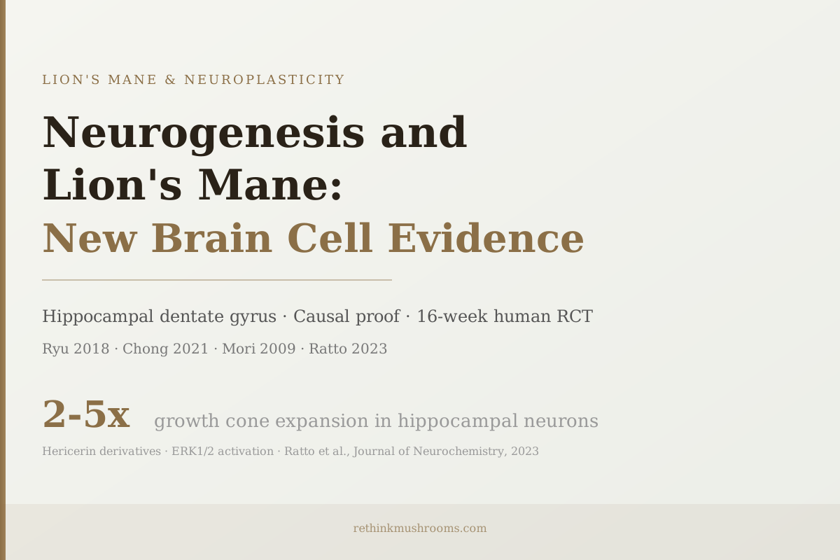
2-5x
growth cone expansion in hippocampal neurons after hericerin derivatives (Ratto et al., 2023)
Causal proof
Chong 2021 showed blocking neurogenesis pharmacologically blocked the antidepressant effect of lion's mane
Mori et al., Phytotherapy Research, 2009
 View as image ↗
Most discussions of lion's mane and the brain stop at nerve growth factor. NGF is the compound lion's mane is famous for stimulating, and the research is real. But NGF is a means to an end. The more specific question is what it leads to: do the neurotrophic compounds in lion's mane actually cause new brain cells to grow? The answer, in adult animals, appears to be yes. The region where new neuron growth matters most for memory and mood, the hippocampal dentate gyrus, is where H. erinaceus extract has been shown to promote neurogenesis. And in a 2021 study, researchers blocked that neurogenesis with a pharmacological inhibitor and showed that blocking it removed the mushroom's antidepressant effect, establishing causality rather than correlation. This article is part of the broader guide to what lion's mane does to the brain.
Key Takeaways
- Adult neurogenesis occurs primarily in the hippocampal dentate gyrus and olfactory bulb; hippocampal neurogenesis specifically drives learning, memory consolidation, and emotional regulation
- Ryu et al. (2018) found H. erinaceus extract specifically promoted neurogenesis in the hippocampus of adult mice AND reduced anxiety and depressive behaviors in the same animals[1]
- Chong et al. (2021) established causality: pharmacologically blocking hippocampal neurogenesis removed lion's mane's antidepressant effect; the benefit was neurogenesis-dependent, not coincidental[2]
- Hericerin derivatives from lion's mane fruiting body activated all four neurotrophins (NGF, BDNF, NT3, NT4/5) in hippocampal neurons, increasing growth cone area by 2-5x, the cellular structures that form new synaptic connections[4]
- Mori et al. (2009) found lion's mane at 3g/day produced significantly higher cognitive scores in MCI patients over 16 weeks; scores declined after supplementation stopped, consistent with neurogenesis requiring ongoing stimulus[3]
- The key honest limitation: hippocampal neurogenesis in adult humans remains scientifically debated; strong animal evidence exists, but direct human neurogenesis evidence has not been established for any compound, including lion's mane
What Neurogenesis Actually Is and Where It Happens in the Adult Brain
Adult neurogenesis means new neurons forming and integrating into existing neural circuits in a brain that has finished developing. For most of the twentieth century, scientists believed the adult brain could not grow new neurons at all. That consensus shifted in the 1990s, and research in rodents established that new neurons are produced in two specific regions throughout adulthood.
The first is the hippocampal dentate gyrus, a structure within the hippocampus, the region of the brain most responsible for converting short-term experiences into long-term memories and for regulating emotional responses. The second is the olfactory bulb. Both regions continuously replace neurons and integrate new cells into their circuits.
Adult neurogenesis primarily occurs in the hippocampal dentate gyrus and the olfactory bulb. Lion's mane specifically promotes hippocampal neurogenesis through BDNF upregulation and neurotrophic signaling, with evidence from adult mouse studies (Ryu et al., 2018) and causal proof that blocking neurogenesis blocks the antidepressant effect (Chong et al., 2021). New hippocampal neurons contribute to pattern separation (the ability to distinguish similar memories), stress resilience, and mood regulation.
BDNF (brain-derived neurotrophic factor) is the primary molecular signal driving hippocampal neurogenesis. When BDNF levels are high, new neurons form and survive. When BDNF is low, neurogenesis slows, hippocampal volume shrinks over time, and both memory function and mood regulation suffer. This is why BDNF is sometimes called the brain's "fertilizer." It is also why the compounds in lion's mane that stimulate BDNF production are specifically relevant to neurogenesis, not just generic neurotrophic effects.
The Core Study: Hippocampal Neurogenesis and Mood in the Same Experiment
Ryu and colleagues (2018) gave adult mice H. erinaceus extract and then measured two things: whether new neurons had formed in the hippocampus, and whether the animals behaved differently in anxiety and depression tests.[1]
Both effects were positive. The extract promoted neurogenesis in the hippocampal dentate gyrus of adult mice. The same animals showed reduced anxiety and depressive behaviors compared to controls.
That both effects occurred together in the same experiment is meaningful. Antidepressants like SSRIs are also associated with hippocampal neurogenesis. The longstanding question in antidepressant research is whether the mood improvement from these drugs is actually a side effect of their neurogenesis promotion, rather than their direct effect on serotonin signaling. That debate remains unresolved. But it frames why a compound that promotes hippocampal neurogenesis would be expected to also affect mood.
The Ryu finding does not by itself prove that neurogenesis caused the mood benefit. Both effects could share a common upstream cause without being directly related. That is where the next study matters.
The Causal Test: Blocking Neurogenesis Blocks the Effect
Chong and colleagues (2021) ran the critical experiment.[2] They used a pharmacological agent to block hippocampal neurogenesis in animals receiving H. erinaceus. The question was: does blocking neurogenesis also block the antidepressant effect?
The answer was yes. When neurogenesis was pharmacologically prevented, lion's mane's antidepressant effect disappeared. This moves the relationship from correlation to causality. The antidepressant effect of H. erinaceus is neurogenesis-dependent, not just neurogenesis-correlated. The mushroom does not produce mood changes through some other pathway that also happens to stimulate neurogenesis. Neurogenesis is the mechanism.
This type of causal proof, often called a "pharmacological blockade" experiment, is standard in neuroscience for establishing that a proposed mechanism is necessary and not just associated. By this standard, Chong 2021 is the mechanistically strongest piece of evidence in the lion's mane neurogenesis literature.
This result matters for the broader discussion of lion's mane and mood in the neuroplasticity overview. The antidepressant and anxiolytic effects found across multiple studies are not separate from the neurogenesis story. They are the functional expression of it.
What Triggers New Neuron Growth: The Molecular Chain
How exactly do lion's mane compounds cause new neurons to grow? The chain involves several steps.
Hericenones, compounds found in the lion's mane fruiting body, and erinacines, found in the mycelium, stimulate the production of neurotrophic factors in the brain. BDNF is the most important for hippocampal neurogenesis specifically; it binds to TrkB receptors on neural stem cells in the dentate gyrus and triggers their differentiation into new neurons.
Ratto and colleagues (2023) established the most detailed picture of how hericenone-derived compounds (hericerins) work at the cellular level.[4] They found that these compounds activate all four neurotrophins simultaneously, NGF, BDNF, NT3, and NT4/5, in hippocampal neurons, a profile the researchers called "pan-neurotrophic." The activation converges on ERK1/2 signaling, a molecular cascade that controls cell growth, proliferation, and differentiation.
One of the most concrete measures in the Ratto 2023 paper was growth cone area. Growth cones are the tips of developing neurons that navigate toward their synaptic targets and form new connections. In hippocampal neurons exposed to hericerin compounds, growth cone area increased by 2 to 5 times. Larger growth cones mean more potential for synaptic connectivity, which is neuroplasticity at its most fundamental cellular level.
For the deeper mechanistic picture on how hericenones enter the brain and stimulate NGF synthesis, hericenones and NGF synthesis: how lion's mane stimulates nerve growth covers the compound-level detail. For erinacines specifically and why they can cross the blood-brain barrier, erinacines: the compounds that cross the blood-brain barrier explains the molecular size difference that makes their brain access possible.
Human Evidence: Cognitive Improvements on a Neurogenesis-Consistent Timeline
Animal neurogenesis studies establish that the mechanism exists. Human clinical data tests whether the functional outcome, improved cognition, actually follows.
Mori and colleagues (2009) conducted the foundational human trial: a double-blind, placebo-controlled RCT in 30 subjects aged 50-80 with mild cognitive impairment (MCI).[3] Participants received lion's mane at 3 grams per day for 16 weeks. Cognitive scores measured by the Revised Hasegawa Dementia Scale were significantly higher in the lion's mane group at weeks 8, 12, and 16 compared to placebo. After supplementation stopped, scores declined.
The 16-week timeline is telling. Hippocampal neurogenesis takes weeks because newly born neurons require time to mature, migrate to their final position, and form functional synaptic connections. The gradual improvement across weeks 8 through 16 in Mori 2009 fits the expected neurogenesis timeline. The score decline after stopping fits the observation that neurogenesis requires ongoing stimulus. When the BDNF-promoting signal stops, the rate of new neuron production slows.
This is not proof that neurogenesis caused the cognitive improvement in humans. That would require a human pharmacological blockade experiment, which is not ethically feasible. But the timeline, the dose-response progression, and the post-treatment decline pattern are all consistent with a neurogenesis-mediated effect.
Docherty and colleagues (2023) extended the human evidence to healthy young adults, a population for whom the question is not cognitive decline but cognitive performance and stress resilience.[6] In their double-blind parallel-groups pilot RCT, chronic lion's mane supplementation produced a trend toward reduced subjective stress over 28 days. Neurogenesis-supporting compounds are not only relevant to MCI populations.
BDNF as a Human Biomarker: Vigna 2019 and What It Measured
Vigna and colleagues (2019) ran the only human study that directly measured circulating BDNF changes following lion's mane supplementation.[5] In overweight and obese patients, supplementation improved mood and sleep disorders, and these improvements were associated with changes in circulating pro-BDNF and BDNF levels.
The measurement requires careful interpretation. Circulating BDNF in the blood is not the same as BDNF in the brain. The blood-brain barrier limits direct transport. Brain BDNF is not measurable in living humans by any currently available method. What Vigna measured was blood-level changes that may reflect altered brain BDNF production but are not a direct measure of it.
The pro-BDNF distinction also matters. Pro-BDNF, the precursor form of BDNF before it is cleaved to its mature form, has different effects on neurons. Mature BDNF binds TrkB receptors and promotes neuronal survival. Pro-BDNF binds p75 neurotrophin receptors and can trigger apoptosis (cell death) in certain contexts. The direction of change in the Vigna study in circulating pro-BDNF, and what that suggests about central BDNF status, is not definitively interpretable from blood levels alone.
What Vigna 2019 does establish: lion's mane supplementation produces measurable changes in BDNF-related biomarkers in humans, associated with improved mood and sleep, consistent with a central neurotrophic effect. It is indirect evidence, but it is human evidence where most of the field has only animal evidence.
Protecting Neurogenesis Under Neurological Threat
Rodriguez and colleagues (2022) tested lion's mane in a tau pathology mouse model (rTg4510), an animal designed to replicate aspects of tau-related neurodegeneration.[7] Even in animals with tau-related neurological pathology, lion's mane produced anxiolytic effects.
This finding extends the neurogenesis conversation from healthy brains to pathologically compromised ones. Tau accumulation impairs hippocampal function and disrupts neuroplasticity. The fact that lion's mane can produce anxiolytic effects in this context suggests that its neuroplasticity-supporting mechanisms function even when the underlying neural environment is compromised.
A comprehensive 2024 review by Cha and colleagues in Neuroscience and Biobehavioral Reviews assessed the evidence for mushrooms and neurocognitive health across the lifespan.[8] The review concluded that while the animal and mechanistic evidence for H. erinaceus neurological effects is substantial, most human trials are small, short-duration, and limited in population diversity. This is an honest summary of the evidence base as of 2024.
For the connection between neurogenesis protection and Alzheimer's disease specifically, lion's mane and amyloid beta: mechanisms of neuroprotection covers how the neuroplasticity-supporting mechanisms may slow the amyloid-related disruption of hippocampal function.
The Honest Uncertainty: What the Human Translation Requires
The most important caveat in the lion's mane neurogenesis literature is one that applies to all neurogenesis research: adult hippocampal neurogenesis in humans is still scientifically debated.
Multiple studies using postmortem human hippocampal tissue have produced contradictory findings. Some groups find evidence of newly formed neurons in the human adult dentate gyrus. Others find no such evidence. The methodological challenges are significant: tissue fixation, antibody specificity, and the time between death and tissue processing all affect the results. The debate remains unresolved.
The lion's mane animal evidence is strong. Hippocampal neurogenesis in adult rodents is well-established, and the Ryu 2018 and Chong 2021 studies demonstrate both the effect and its causal necessity. The human clinical trial data (Mori 2009, Docherty 2023, Vigna 2019) shows functional cognitive and mood benefits consistent with a neurogenesis hypothesis. But none of these human studies directly measure neurogenesis.
The honest position: lion's mane promotes adult hippocampal neurogenesis in animal models, through a mechanism with solid causal evidence (Chong 2021). Human clinical trials show functional improvements consistent with neurogenesis timelines. Direct human neurogenesis evidence does not yet exist for lion's mane or for any other compound. The mechanistic case is compelling; the human translation is supported but not proven.
For the broader picture of neuroplasticity mechanisms including synaptic plasticity and long-term potentiation alongside neurogenesis, how lion's mane supports synaptic plasticity covers the complementary cellular mechanisms.
The Bottom Line
Lion's mane promotes adult hippocampal neurogenesis in animal studies, with Ryu et al. (2018) demonstrating the effect and Chong et al. (2021) establishing causality by showing that pharmacological blockade of neurogenesis removes the antidepressant benefit. Hericerin derivatives activate all four neurotrophins via ERK1/2 signaling, expanding growth cone area 2-5x (Ratto et al., 2023). Human clinical data shows cognitive improvements on a 16-week neurogenesis-consistent timeline (Mori 2009) and BDNF biomarker changes in humans (Vigna 2019). Adult human hippocampal neurogenesis remains scientifically debated; direct human evidence does not yet exist.
Frequently Asked Questions
Sources & References
- 1. Source details for this reference are not yet available.
- 2. Source details for this reference are not yet available.
- 3. Source details for this reference are not yet available.
- 4. Source details for this reference are not yet available.
- 5. Source details for this reference are not yet available.
- 6. Source details for this reference are not yet available.
- 7. Source details for this reference are not yet available.
- 8. Source details for this reference are not yet available.