Agaricus blazei and Immune Function
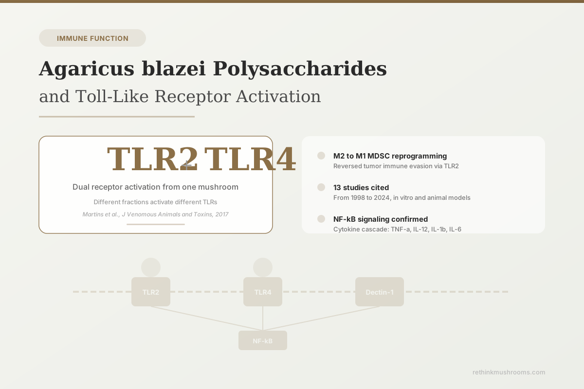
TLR2 + TLR4
Agaricus blazei Murill polysaccharides activate at least two distinct toll-like receptors on human immune cells, each triggering different downstream cytokine profiles and functional outcomes.
Martins et al., J Venomous Animals and Toxins, 2017
NF-kB Activation
confirmed via TLR2 in human monocytes
M2 to M1 Shift
tumor-suppressive MDSC reprogramming
No Human Trials
all TLR data from in vitro and animal models
Martins et al., J Venomous Animals and Toxins, 2017
 View as image ↗
Your immune system evolved to detect fungal polysaccharides. The pattern recognition receptors on macrophages, dendritic cells, and monocytes treat beta-glucans from mushroom cell walls as signals of potential invasion. Agaricus blazei Murill (ABM), also known as Agaricus brasiliensis, contains polysaccharide complexes that activate at least two of these receptors: toll-like receptor 2 (TLR2) and toll-like receptor 4 (TLR4). The activation is not generic. Different polysaccharide fractions from the same mushroom trigger different receptors, produce different cytokine profiles, and drive different functional outcomes in immune cells. This specificity matters because it determines whether the immune response tilts toward inflammation, anti-tumor activity, or pathogen clearance. For the broader landscape of ABM bioactive compounds, including non-polysaccharide components like blazein, a separate article covers the full chemical profile.
Key Takeaways
- ABM polysaccharide (pAbM) acted as a specific TLR2 agonist in mice, reprogramming immunosuppressive M2-type myeloid-derived suppressor cells into pro-inflammatory M1 cells; TLR2-deficient mice showed no response (Liu et al., Immunology, 2015)
- An acid-treated polysaccharide fraction from A. brasiliensis significantly increased TLR2 and TLR4 expression on human monocytes and enhanced phagocytosis of Candida albicans (Martins et al., J Venomous Animals and Toxins, 2017)
- ABM fraction H (ABH) induced IL-12 production in human monocytes via CD14/TLR4, not TLR2; anti-TLR4 antibody blocked the response in a dose-dependent manner (Kasai et al., Evidence-Based CAM, 2004)
- Cold water extract from A. brasiliensis bound directly to TLR2 protein, bypassing Dectin-1 entirely because the beta-glucan segments were too small for Dectin-1 recognition (Yamanaka et al., Immunopharmacol Immunotoxicol, 2012)
- ABM polysaccharide significantly promoted CD8+ T cell activity against colorectal cancer cells in co-culture and inhibited intraperitoneal tumor growth in mice (Xu et al., Int J Biol Macromolecules, 2024)
- Beta-glucans from Agaricus species drove trained immunity in human monocytes, reprogramming metabolic and epigenetic pathways for enhanced cytokine responses to secondary stimulation (Case et al., Frontiers in Nutrition, 2024)
What Toll-Like Receptors Do
Toll-like receptors (TLRs) are a family of pattern recognition receptors that sit on the surface of immune cells. They evolved to detect conserved molecular structures from pathogens: bacterial lipopolysaccharide (TLR4), bacterial lipoproteins (TLR2), viral double-stranded RNA (TLR3), and fungal cell wall components (TLR2, TLR4, and others). Humans have 10 functional TLRs, each recognizing a distinct set of molecular patterns.
When a TLR binds its ligand, it triggers an intracellular signaling cascade. The canonical pathway runs through the adaptor protein MyD88 to NF-kB, a transcription factor that turns on genes for inflammatory cytokines (TNF-alpha, IL-1beta, IL-6, IL-12), chemokines, and co-stimulatory molecules. This is the bridge between innate and adaptive immunity: TLR activation on dendritic cells and macrophages determines how T cells and B cells are later instructed to respond. For a deeper look at how this leads to NK cell activation, the NK cell article covers the downstream consequences for cytotoxic lymphocytes.
Two TLRs are particularly relevant to ABM polysaccharides:
- TLR2 forms heterodimers with TLR1 or TLR6 and recognizes a broad range of microbial products, including fungal polysaccharides. It signals exclusively through MyD88.
- TLR4 is best known as the receptor for bacterial LPS. It can signal through both MyD88 and a second adaptor called TRIF, giving it access to a wider range of downstream pathways, including type I interferon production.
TLR2: The Primary ABM Receptor
The strongest evidence for TLR2 as a primary ABM receptor comes from Liu and colleagues (2015), who demonstrated that polysaccharide ABM (pAbM) is a specific TLR2 agonist.[1] Their experiments showed that pAbM reprogrammed myeloid-derived suppressor cells (MDSCs) from an immunosuppressive M2 phenotype to a pro-inflammatory M1 phenotype. The critical evidence for TLR2 specificity: pAbM had no effect in TLR2-deficient mice. The entire anti-tumor response required intact TLR2 signaling.
Functionally, TLR2-activated MDSCs showed:
- Increased IL-12, TNF-alpha, and iNOS expression
- Decreased Arginase 1, TGF-beta, and IL-10 expression
- Reduced ability to suppress T cell proliferation
- Reduced conversion of CD4+ T cells into regulatory T cells
This M2-to-M1 shift is significant because MDSCs are one of the primary mechanisms tumors use to evade immune detection. By switching these cells from immunosuppressive to immunostimulatory through TLR2, ABM polysaccharides reversed the tumor's immune evasion strategy in this mouse model.
Yamanaka and colleagues (2012) provided a different angle on TLR2 activation.[2] They prepared a cold water extract from A. brasiliensis fruit bodies and found that the 1,3-beta-glucan segments in this fraction were too small to interact with Dectin-1, the primary beta-glucan receptor. Despite bypassing Dectin-1 entirely, the extract still stimulated cytokine production. Through systematic blocking studies, they demonstrated that TLR2 was the responsible receptor. The extract bound directly to TLR2 protein, and anti-TLR2 monoclonal antibody suppressed IL-6 production from splenocytes.
This finding matters because it shows ABM contains water-soluble TLR2 ligands that are distinct from the large beta-glucans typically credited with immunomodulation. The molecules responsible are smaller polysaccharide complexes or non-glucan components that activate a different receptor pathway entirely.
TLR4: The Alternative Recognition Route
Not all ABM fractions activate TLR2. Kasai and colleagues (2004) isolated a specific fraction called ABH from ABM and tested its ability to induce IL-12 production in human monocytes and macrophages.[3] IL-12 is a critical cytokine for driving Th1 immune responses, which favor cellular immunity and anti-tumor activity.
ABH-induced IL-12 production was:
- Blocked by anti-CD14 and anti-TLR4 antibodies in a dose-dependent manner
- Not blocked by anti-TLR2 antibodies
- Not inhibited by polymyxin B, ruling out endotoxin contamination as the cause
The CD14/TLR4 dependency mirrors the signaling pathway used by bacterial lipopolysaccharide, but the polymyxin B resistance confirms that ABH is not simply contaminated with LPS. ABH contains a polysaccharide component that is recognized by TLR4 through a mechanism distinct from endotoxin recognition. In vivo, oral ABH enhanced NK cell activity in mouse splenocytes, connecting TLR4 activation to downstream cytotoxic function.
Martins and colleagues (2017) showed that both receptors can be activated simultaneously.[4] An acid-treated polysaccharide fraction (ATF) from A. brasiliensis significantly increased expression of both TLR2 and TLR4 on human monocytes. The functional consequences differed by receptor:
- TLR4 activation was associated with higher TNF-alpha production
- TLR2 activation was associated with higher IL-1beta production
- Blocking either receptor substantially reduced monocyte phagocytosis of Candida albicans
IL-10 production increased independently of TLR activation, suggesting additional receptor pathways (possibly Dectin-1 or complement receptor 3) contribute to the full immunomodulatory profile. The co-activation of TLR2 and TLR4 from a single polysaccharide fraction distinguishes ABM from simpler immunostimulants that target only one receptor.
Why Different Fractions Activate Different Receptors
The structural diversity within ABM polysaccharides explains the receptor selectivity. Smiderle and colleagues (2011) compared polysaccharides from A. brasiliensis and A. bisporus (the common button mushroom) and found structural similarities in their beta-glucan backbones but differences in branching patterns and fucose content.[5] Both species' polysaccharides activated NF-kB in human THP-1 monocytes, but the magnitude and cytokine profile differed.
For the detailed structural analysis of 1,3/1,6 beta-D-glucan, a dedicated article breaks down how branching patterns determine receptor binding. The key principle: TLR2 recognizes smaller, water-soluble polysaccharide complexes and possibly non-glucan components, while TLR4 responds to larger polysaccharide fractions that may contain fucose or other structural modifications. Dectin-1, the third major receptor, requires beta-1,3-glucan chains of sufficient length to form the triple-helix structure needed for binding.
This means extraction method determines which receptors a given ABM preparation activates. Hot water extraction yields larger polysaccharides that may favor TLR4 and Dectin-1. Cold water extraction captures smaller molecules that preferentially activate TLR2. Acid treatment exposes different epitopes and can activate both. The extraction variables explain why different research groups report different primary receptors for what is nominally the same mushroom.
Downstream Signaling: From Receptor to Response
Bernardshaw and colleagues (2005) mapped the downstream consequences of ABM-mediated TLR activation in human cells.[6] ABM extract differentially stimulated pro-inflammatory cytokines in human monocytes versus human vein endothelial cells (HUVECs). Monocytes produced large amounts of TNF-alpha, IL-1beta, and MCP-1. Endothelial cells produced different cytokine profiles despite expressing the same TLRs, demonstrating that the cellular context determines the outcome of TLR activation.
The NF-kB pathway is central to all of these responses. When TLR2 or TLR4 binds its ligand on an ABM polysaccharide, MyD88 is recruited to the receptor's intracellular domain. MyD88 activates IRAK kinases, which phosphorylate TRAF6, which ultimately activates the IKK complex. IKK phosphorylates IkB, releasing NF-kB to translocate to the nucleus and turn on inflammatory gene expression. Liu and colleagues (2017) confirmed this pathway in vivo: ABM polysaccharides modulated TLR4 expression, NF-kB activation, and downstream cytokine levels (IL-1beta, IL-6, TNF-alpha) in chicken lymphocytes.[7]
The cytokine profile driven by TLR activation determines the direction of the adaptive immune response:
- IL-12 (TLR4-driven): pushes naive T cells toward Th1 differentiation, favoring cellular immunity and anti-tumor responses
- TNF-alpha (both TLR2 and TLR4): activates macrophages, promotes inflammation, enhances antigen presentation
- IL-1beta (TLR2-driven): co-stimulates T cell activation, enhances dendritic cell maturation
- IL-6 (both pathways): drives B cell differentiation into antibody-producing plasma cells
This pathway explains how ABM polysaccharides connect to the vaccine adjuvant research: TLR activation on dendritic cells provides the co-stimulatory signals needed for robust adaptive immune responses.
Tumor Immunity: MDSC Reprogramming and CD8+ T Cells
The tumor immunology applications of ABM TLR activation are built on two mechanisms. The first, described above, is the TLR2-dependent reprogramming of MDSCs from M2 to M1.[1] The second comes from Xu and colleagues (2024), who tested ABM polysaccharide (ABMP) against intraperitoneal metastatic colorectal cancer in mice.[8]
In vitro, ABMP significantly promoted CD8+ T cell activity and enhanced their colorectal cancer cell killing function. In vivo, oral ABMP inhibited the growth of intraperitoneal tumors in mice when given as a preventive intervention. The mechanism connected to both immune cell activation and changes in the gut microbiota, suggesting that TLR-mediated effects in the intestinal immune compartment contribute to systemic anti-tumor responses.
These findings are preclinical. No human cancer trial has tested ABM polysaccharides with TLR activation as a defined endpoint. But the mechanistic chain from TLR activation to MDSC reprogramming to enhanced cytotoxic T cell function provides a coherent biological pathway for the anti-tumor effects reported in Agaricus blazei cancer research.
Gut Immunity and Intestinal TLR Activation
TLR activation is not limited to injected or intravenous polysaccharides. The intestinal epithelium expresses TLR2 and TLR4, and orally administered ABM polysaccharides interact with these receptors in the gut-associated lymphoid tissue (GALT).
Ji and colleagues (2023) demonstrated that ABM polysaccharide alleviated DSS-induced colitis in mice by modulating the intestinal barrier and remodeling metabolism.[9] Zhao and colleagues (2024) showed that ABM polysaccharide protected against intestinal ischemia/reperfusion injury through regulating gut microbiota composition and reducing inflammatory cytokine levels.[10]
In the context of gut immunity, TLR activation by ABM polysaccharides appears to follow a different pattern than systemic activation. Rather than driving inflammation, intestinal TLR engagement by dietary polysaccharides can reinforce barrier function, support commensal bacteria, and reduce pathological inflammation. This paradox, where the same receptors produce opposing outcomes depending on context, reflects the tissue-specific programming of TLR signaling in the gut versus the blood.
Li and colleagues (2019) provided further evidence that ABM polysaccharides modulate TLR4 pathway activity in immune organs, reducing oxidative damage and inflammatory cytokine levels in a cadmium toxicity model.[11] The protective effect was associated with decreased TLR4 expression and downstream NF-kB activity in the spleen and thymus, demonstrating that ABM polysaccharides can either activate or dampen TLR signaling depending on the inflammatory context.
Trained Immunity: Epigenetic Reprogramming
Case and colleagues (2024) added a new dimension to ABM polysaccharide immunology: trained immunity.[12] Trained immunity is a form of innate immune memory where monocytes and macrophages, after initial stimulation with beta-glucans, show enhanced responses to unrelated secondary stimuli days or weeks later. This is not classical adaptive immunity (no T or B cell memory). Instead, it involves metabolic and epigenetic reprogramming of innate immune cells.
Beta-glucans from Agaricus species drove trained immunity in human monocytes, increasing their capacity for cytokine production upon restimulation. The mechanism involves histone modifications (H3K4 methylation, H3K27 acetylation) at promoter regions of inflammatory genes, combined with a metabolic shift from oxidative phosphorylation toward aerobic glycolysis.
TLR signaling is the entry point for this reprogramming. The initial beta-glucan exposure activates TLR2 and Dectin-1, which trigger the downstream signaling that ultimately modifies the epigenetic landscape of the cell. The trained state persists after the original stimulus is cleared, because the epigenetic marks are heritable across cell divisions. For the Th1/Th2 balance implications of this immune reprogramming, the allergy article covers how trained innate immunity intersects with adaptive immune polarization.
Limitations of Current Evidence
Several constraints apply to the TLR-ABM research:
The receptor identification studies rely heavily on blocking antibodies and knockout mice. These tools are imperfect. Anti-TLR2 antibodies may not fully block all TLR2 signaling, and knockout mice have developmental compensations that wild-type organisms lack. Direct binding studies (like Yamanaka's TLR2 binding data) are stronger evidence, but only one group has published direct binding data.
Extraction method determines which polysaccharide fractions are present, which in turn determines which TLRs are activated. This makes it difficult to compare results across studies using different extraction protocols. A hot water extract, a cold water extract, and an acid-treated fraction from the same mushroom can activate different receptors and produce different cytokine profiles. Standardization of extraction methods has not been achieved.
The cadmium toxicity models (Liu 2017, Li 2019) use an artificial stress context. Whether TLR modulation under heavy metal exposure translates to normal physiological conditions is unclear.
No human study has measured TLR expression or activation as a primary endpoint after ABM supplementation. The Mizuno (1998) study showing increased T cell subsets in mice after oral ABM polysaccharides[13] is consistent with TLR-mediated immune activation, but did not measure TLR signaling directly. The gap between mechanistic in vitro data and clinical relevance remains substantial. Whether TLR-mediated immune activation translates to benefit for immune-compromised patients is a separate question explored in its own article. For a comparative view of where ABM sits relative to other species, the article examining whether ABM is the most immunostimulating mushroom puts these TLR findings in a cross-species context.
The Bottom Line
Agaricus blazei Murill polysaccharides activate toll-like receptors 2 and 4 on immune cells, triggering NF-kB signaling, cytokine production, and functional changes including MDSC reprogramming and enhanced phagocytosis. Which receptor is activated depends on the polysaccharide fraction and extraction method. All evidence comes from in vitro experiments and animal models. No human trial has directly measured TLR activation from ABM supplementation.
Frequently Asked Questions
Sources & References
- 1RTHM-00395·Liu, Yi et al. (2015). “Polysaccharide Agaricus blazei Murill stimulates myeloid derived suppressor cell differentiation from M2 to M1 type, which mediates inhibition of tumour immune-evasion via the Toll-like receptor 2 pathway..” Immunology.Study breakdown →PubMed →↩
- 2RTHM-00273·Yamanaka, Daisuke et al. (2012). “Effect of Agaricus brasiliensis-derived cold water extract on Toll-like receptor 2-dependent cytokine production in vitro..” Immunopharmacology and immunotoxicology.Study breakdown →PubMed →↩
- 3RTHM-00071·Kasai, H et al. (2004). “IL-12 Production Induced by Agaricus blazei Fraction H (ABH) Involves Toll-like Receptor (TLR)..” Evidence-based complementary and alternative medicine : eCAM.Study breakdown →PubMed →↩
- 4RTHM-00483·Martins, Priscila Raquel et al. (2017). “Agaricus brasiliensis polysaccharides stimulate human monocytes to capture Candida albicans, express toll-like receptors 2 and 4, and produce pro-inflammatory cytokines..” The journal of venomous animals and toxins including tropical diseases.Study breakdown →PubMed →↩
- 5RTHM-00234·Smiderle, Fhernanda R et al. (2011). “Polysaccharides from Agaricus bisporus and Agaricus brasiliensis show similarities in their structures and their immunomodulatory effects on human monocytic THP-1 cells..” BMC complementary and alternative medicine.Study breakdown →PubMed →↩
- 6RTHM-00078·Bernardshaw, Soosaipillai et al. (2005). “An extract of the medicinal mushroom Agaricus blazei Murill differentially stimulates production of pro-inflammatory cytokines in human monocytes and human vein endothelial cells in vitro..” Inflammation.Study breakdown →PubMed →↩
- 7RTHM-00482·Liu, Wenjing et al. (2017). “The Effects of Agaricus blazei Murill Polysaccharides on Cadmium-Induced Apoptosis and the TLR4 Signaling Pathway of Peripheral Blood Lymphocytes in Chicken..” Biological trace element research.Study breakdown →PubMed →↩
- 8RTHM-00916·Xu, Yongzhao et al. (2024). “Preventive intervention with Agaricus blazei murill polysaccharide exerts anti-tumor immune effect on intraperitoneal metastasis colorectal cancer..” International journal of biological macromolecules.Study breakdown →PubMed →↩
- 9RTHM-00831·Ji, Zhong-Hao et al. (2023). “Agaricus blazei Polysaccharide Alleviates DSS-Induced Colitis in Mice by Modulating Intestinal Barrier and Remodeling Metabolism..” Nutrients.Study breakdown →PubMed →↩
- 10RTHM-00919·Zhao, Meiqi et al. (2024). “Polysaccharide Isolated from Agaricus blazei Murill Alleviates Intestinal Ischemia/Reperfusion Injury through Regulating Gut Microbiota and Mitigating Inflammation in Mice..” Journal of agricultural and food chemistry.Study breakdown →PubMed →↩
- 11RTHM-00569·Li, Ruyue et al. (2019). “Effects of Fungal Polysaccharide on Oxidative Damage and TLR4 Pathway to the Central Immune Organs in Cadmium Intoxication in Chickens..” Biological trace element research.Study breakdown →PubMed →↩
- 12RTHM-00867·Case, Sarah et al. (2024). “β-glucans from Agaricus bisporus mushroom products drive Trained Immunity..” Frontiers in nutrition.Study breakdown →PubMed →↩
- 13RTHM-00026·Mizuno, M et al. (1998). “Polysaccharides from Agaricus blazei stimulate lymphocyte T-cell subsets in mice..” Bioscience.Study breakdown →PubMed →↩