Ergothioneine & Pregnancy
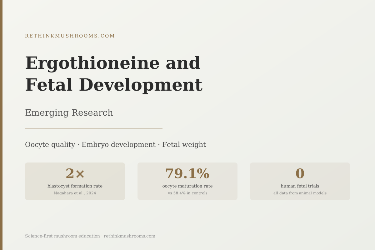
2x blastocyst formation rate
Ergothioneine at 10 μM more than doubled successful blastocyst development in porcine embryos, from 7.4% to 18.9%, while lifting oocyte maturation from 58% to 79%.
Nagahara et al., Veterinary World, 2024
79.1%
oocyte maturation rate vs 58.4% in controls
25 mg/kg/day
dose that rescued fetal weight in rat preeclampsia model
0
human fetal development trials completed to date
Nagahara et al., Veterinary World, 2024
 View as image ↗
In 2020, Williamson and colleagues gave pregnant rats with a surgically induced form of preeclampsia a daily dose of ergothioneine. The treated rats showed significantly better fetal weights at delivery compared to untreated rats with the same condition.[1] The ergothioneine-treated animals also had lower blood pressure and fewer markers of placental distress. It was not a cure, but it was a measurable improvement in an animal model designed to mimic one of the most common causes of poor fetal outcomes in humans.
That study raised a question researchers are still working to answer: does ergothioneine, the sulfur-containing antioxidant found almost exclusively in mushrooms, influence fetal development beyond the preeclampsia context? A small but growing body of animal and laboratory research suggests it might, spanning three distinct windows: the quality of the egg before fertilization, the health of the embryo in its earliest days, and the growth of the fetus in late gestation. None of these findings come from human trials, and that gap matters enormously. But the evidence is specific enough to be worth examining carefully.
Ergothioneine is often called a longevity antioxidant, but its role in reproduction and development is less discussed. For a fuller picture of what it is and how the body acquires it, the ergothioneine overview article covers the biochemistry and dietary sources in detail.
Key Takeaways
- In a 2024 porcine study, ergothioneine at 10 μM raised oocyte maturation rates from 58.4% to 79.1% and more than doubled blastocyst formation from 7.4% to 18.9%
- A 2002 rat study found ergothioneine reduced embryo malformation rates in diabetic pregnant animals to levels comparable to healthy controls, with additive protection alongside vitamin E
- The OCTN1 transporter (SLC22A4) is expressed in the placenta, providing a documented biological mechanism for maternal-to-fetal ergothioneine transfer
- In a rat preeclampsia model, 25 mg/kg/day ergothioneine significantly rescued fetal weight alongside reductions in blood pressure and the antiangiogenic marker sFlt-1
- The common thread across all three developmental windows is oxidative stress: ergothioneine scavenges the reactive oxygen species that disrupt oocyte maturation, embryo formation, and placental function
- Every data point reviewed here is from animal or in vitro research; no human fetal development trials have been completed
Oocyte Quality: Where Reproduction Begins
Before any fetal development can occur, an egg must mature successfully. Oocyte quality, how well the egg reaches its final pre-fertilization state, turns out to be sensitive to oxidative stress. When reactive oxygen species accumulate in the follicular environment, they damage the egg's DNA, disrupt the spindle apparatus that separates chromosomes during division, and lower the chances of successful fertilization and implantation.
In 2024, Nagahara and colleagues added ergothioneine to porcine oocyte maturation media at a concentration of 10 μM and tracked results through the full maturation process.[2] Metaphase II oocyte maturation, the measure of eggs that reached the final developmentally competent state, rose from 58.4% in controls to 79.1% with ergothioneine treatment. DNA fragmentation dropped. When the researchers tracked fertilized eggs forward to the blastocyst stage, the hollow ball of cells that implants in the uterine wall, blastocyst formation rate climbed from 7.4% to 18.9%.
That last number is worth pausing on. Blastocyst formation is one of the most stringent filters in early reproduction. Many fertilized eggs fail to reach this stage, and anything that more than doubles the rate is not producing a marginal effect.
Nagahara's model used pig eggs, which share meaningful biological similarities with human reproductive physiology, but extrapolating directly to human fertility requires caution. These were in vitro results under controlled conditions. Whether ergothioneine from dietary mushroom intake would produce equivalent oocyte benefits in human women depends on baseline ergothioneine status, individual oxidative load, and many other factors the study could not address.
What the study does establish clearly is that ergothioneine has direct biological effects on egg cell quality at concentrations achievable through dietary intake. The proposed mechanism is antioxidant protection during the window when the developing oocyte is most vulnerable to oxidative damage.
Diabetic Pregnancy and the Embryo Malformation Question
One of the most specific findings in this space came from Guijarro and colleagues in 2002 and has received almost no attention in the two decades since.[3]
The experiment was straightforward. Diabetic pregnant rats, whose offspring have elevated rates of structural malformations, were given ergothioneine supplementation during the critical window when organ systems form. Malformation rates dropped to levels comparable to non-diabetic controls. When ergothioneine was combined with vitamin E, the protective effect was additive.
Guijarro's interpretation was that reactive oxygen species from maternal diabetes were the proximate cause of the malformations, and that ergothioneine interrupted this pathway. Diabetic pregnancy generates a substantially higher oxidative burden in the embryonic environment, and the embryo at the stage of organogenesis is especially sensitive to oxidative damage. Ergothioneine appeared to provide a protective buffer against this specific chemical stressor.
This matters beyond the diabetic pregnancy context because oxidative stress during early embryonic development has been linked to structural malformations, neural tube defects, and implantation failure across multiple pregnancy models. If ergothioneine can interrupt this pathway in one oxidative context, the mechanism may apply more broadly, though this has not been tested.
The absence of follow-up research is notable. No human study has tested ergothioneine supplementation during the embryonic period, and no subsequent animal studies appear to have extended Guijarro 2002 specifically. It remains a single study on an important question that has not generated the follow-up it merits.
How Ergothioneine Reaches the Fetus: The Transporter Story
A central question about any potential fetal benefit is whether a substance can actually cross from mother to fetus in meaningful amounts. For ergothioneine, the answer involves a protein called OCTN1, encoded by the gene SLC22A4.
In 2005, Gründemann and colleagues published the discovery that OCTN1 is not a general transporter but a dedicated, high-affinity transporter for ergothioneine specifically.[4] The protein has no other known physiological substrate in humans. This specificity is biologically significant; the body has invested in dedicated molecular machinery to move ergothioneine around selectively, which implies ergothioneine has functions important enough to warrant that investment.
Critically, SLC22A4 is expressed in the placenta. A 2020 review by Borodina and colleagues confirmed this distribution in a comprehensive survey of ergothioneine transporter biology, noting that ergothioneine accumulates preferentially in tissues with high oxidative stress exposure, including the liver, kidneys, and red blood cells.[5] These are tissues where mitochondrial activity is highest and oxidative damage is a constant challenge.
The implication is that the placenta is not a passive barrier that ergothioneine happens to cross. The OCTN1 transporter is there for a reason: the placenta actively imports ergothioneine from maternal circulation, and from there it can enter fetal circulation. For a deeper look at OCTN1 biology and what its placental expression tells us, the OCTN1 transporter article covers the molecular detail.
What remains unknown is whether maternal dietary ergothioneine intake translates into clinically meaningful fetal plasma concentrations in humans. The placental transporter provides the biological mechanism; the magnitude of fetal exposure from dietary sources has not been measured in human studies.
For the broader picture of how ergothioneine behaves specifically at the placenta during pregnancy, the placental health research article examines the maternal-fetal biology in depth.
Late-Gestation Fetal Weight: What the Preeclampsia Model Shows
The most clinically relevant animal fetal data comes from Williamson's 2020 study using the reduced uterine perfusion pressure (RUPP) rat model, a standard preclinical tool for preeclampsia research.[1] In this model, blood flow to the uterus is surgically restricted to create a hypertensive pregnancy state that mirrors several features of human preeclampsia.
Rats with RUPP preeclampsia given 25 mg/kg/day ergothioneine showed several improvements compared to untreated RUPP controls: blood pressure was lower, circulating sFlt-1 dropped substantially, and fetal weights were significantly higher. sFlt-1 is a protein released by a stressed placenta that blocks the formation of new blood vessels; its elevation in maternal blood is a recognized marker of preeclampsia severity, and its reduction in the ergothioneine-treated animals suggests the treatment was acting at a mechanistic level rather than masking surface symptoms.
The placental tissue showed upregulation of four proteins central to antioxidant defense: Nrf2 (the body's master antioxidant transcription factor), SOD-1 (the enzyme that converts the most common reactive oxygen species into a less harmful form), PGC-1α (which regulates mitochondrial biogenesis and energy production), and UCP-1 (which reduces mitochondrial heat production and the associated generation of free radicals). Mitochondrial reactive oxygen species in placental cells were measurably lower in ergothioneine-treated animals.
This is mechanistic evidence, not correlation. Supplementation turned on the antioxidant machinery in the placenta, reduced the chemical signature of placental distress, and produced better fetal weights. The animal model is an imperfect proxy for human preeclampsia. But the biological pathway it illuminates, placental oxidative stress driving both hypertension and fetal growth restriction, is the same pathway that operates in human disease.
For the evidence on ergothioneine levels in women who develop preeclampsia, the preeclampsia risk article covers the human cohort findings.
The Common Thread: Why All Three Windows Connect to Oxidative Stress
The developmental arc from egg to embryo to late-gestation fetus looks like three separate research questions, but a single mechanism runs through all of them.
Oocyte maturation is disrupted by reactive oxygen species accumulating in follicular fluid. Embryo malformation in diabetic pregnancy is driven by the oxidative burden created by maternal hyperglycemia. Placental dysfunction in preeclampsia generates mitochondrial reactive oxygen species that impair vascular signaling and restrict nutrient delivery to the fetus. In each case, the fundamental problem is too much oxidative damage, too little capacity to neutralize it.
Ergothioneine operates at the intersection of all three. Kerley and colleagues' 2018 systematic review established this framework in detail, describing ergothioneine's distinguishing mechanism: unlike common dietary antioxidants, it targets mitochondrial reactive oxygen species specifically, activates the Nrf2 pathway and triggers the production of multiple endogenous antioxidant enzymes simultaneously, and accumulates in the tissues (placenta, red blood cells, organs with high metabolic demand) where oxidative stress is most concentrated.[6]
Whether ergothioneine as a dietary compound can shift oxidative status enough to produce clinically meaningful effects during human pregnancy is unanswered. The animal data suggests the mechanism is real and biologically coherent. The dose at which it becomes relevant in humans, and whether that dose is achievable through mushroom consumption, is not established.
The Evidence Limit: Everything Here Comes From Animals
This article examines six studies. Every one is either an animal model or an in vitro experiment. No human fetal development trial has been completed using ergothioneine.
This is not a minor caveat. Animal-to-human translation in reproductive biology is unreliable. Many antioxidant interventions that worked in rodent pregnancy models failed to produce benefits in human trials, and some showed harm. The unique biology of human placentation, the different pharmacokinetics of ergothioneine across species, and the complex interplay of maternal health and genetics mean that nothing in this article should inform clinical decisions.
What this research does establish:
- Ergothioneine has biological activity in reproductive systems across multiple species at each of three developmental windows
- The mechanisms it activates (Nrf2, mitochondrial antioxidant enzymes) are the same mechanisms relevant to human fetal development
- The placenta has the dedicated transporter to receive ergothioneine from maternal blood
- The doses used in animal studies have no confirmed human equivalent
The European Food Safety Authority reviewed ergothioneine safety for pregnant and breastfeeding women and concluded that a sufficient margin of exposure exists at anticipated dietary intake levels. This provides reassurance that culinary mushrooms as food are safe during pregnancy, though it is not evidence of fetal benefit.
What is missing is a human intervention trial: randomized, with pregnant women, measuring fetal outcomes against a placebo. The data that would allow confident conclusions about human fetal development simply does not yet exist. The evidence here is a starting point for that research.
The Bottom Line
Animal research shows ergothioneine acts across three windows of reproductive development: improving oocyte maturation quality, reducing embryo malformation rates in a diabetic pregnancy model, and rescuing fetal weight in a preeclampsia model. The OCTN1 transporter expressed in the placenta provides a documented biological mechanism for maternal-fetal transfer. No human fetal development trials have been completed, and all current findings should be understood as preclinical starting points, not clinical guidance.
Frequently Asked Questions
Sources & References
- 1RTHM-01301·Williamson, Rachel D et al. (2020). “L-(+)-Ergothioneine Significantly Improves the Clinical Characteristics of Preeclampsia in the Reduced Uterine Perfusion Pressure Rat Model..” Hypertension (Dallas.Study breakdown →PubMed →↩
- 2RTHM-01953·Nagahara, Megumi et al. (2024). “Effects of ergothioneine supplementation on meiotic competence and porcine oocyte development..” Veterinary world.Study breakdown →PubMed →↩
- 3RTHM-00090·Guijarro, M V et al. (2002). “Effects of ergothioneine on diabetic embryopathy in pregnant rats..” Food and chemical toxicology : an international journal published for the British Industrial Biological Research Association.Study breakdown →PubMed →↩
- 4RTHM-00154·Gründemann, Dirk et al. (2005). “Discovery of the ergothioneine transporter..” Proceedings of the National Academy of Sciences of the United States of America.Study breakdown →PubMed →↩
- 5RTHM-01175·Borodina, Irina et al. (2020). “The biology of ergothioneine, an antioxidant nutraceutical..” Nutrition research reviews.Study breakdown →PubMed →↩
- 6RTHM-00968·Kerley, Robert N et al. (2018). “The potential therapeutic effects of ergothioneine in pre-eclampsia..” Free radical biology & medicine.Study breakdown →PubMed →↩