Ergothioneine Antioxidant Biology & Mitochondrial Protection
2025
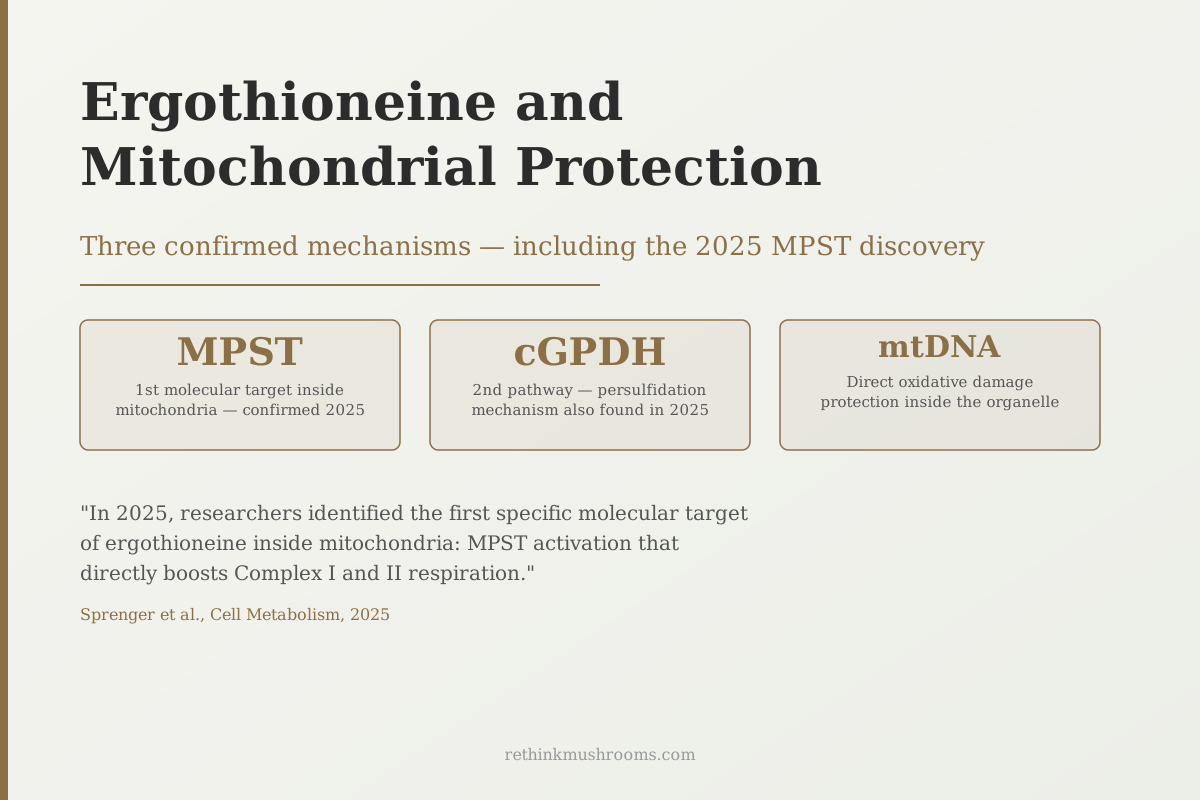
Researchers publishing in Cell Metabolism identified the first specific molecular target of ergothioneine inside mitochondria, an enzyme called MPST that directly boosts cellular respiration.
Sprenger et al., Cell Metabolism, 2025
MPST
first confirmed mitochondrial enzyme target of ergothioneine, identified 2025
2 pathways
distinct mitochondrial mechanisms discovered in 2025 alone (MPST and cGPDH)
40+ years
since Aruoma 1983 first showed ergothioneine outperforms glutathione in mitochondrial membranes
Sprenger et al., Cell Metabolism, 2025
 View as image ↗
In 2025, a team publishing in Cell Metabolism identified the first specific molecular target of ergothioneine inside mitochondria: an enzyme called MPST (3-mercaptopyruvate sulfurtransferase).[1] That finding transformed ergothioneine from a vague antioxidant with claimed mitochondrial benefits into a compound with a known mechanism, one that directly switches on the machinery cells use to generate energy.
Ergothioneine is a sulfur-containing amino acid found almost exclusively in mushrooms. For a broader introduction to what it is and why the human body evolved unusual machinery to accumulate it, see what ergothioneine is and why the body hoards it. This article covers the mitochondrial story specifically: how ergothioneine enters these organelles, what it does once there, and what the evidence actually supports about health relevance.
Key Takeaways
- A 2025 Cell Metabolism study identified MPST as the first confirmed mitochondrial enzyme target of ergothioneine, showing it directly boosts Complex I and Complex II respiration
- Mitochondria take up ergothioneine without needing the OCTN1 transporter that governs ergothioneine uptake at the cell membrane, pointing to a secondary mitochondrial entry route
- A second, completely separate 2025 paper found ergothioneine stimulates cGPDH activity through a persulfidation pathway, improving the electron shuttle that feeds the mitochondrial respiratory chain
- Ergothioneine supplementation in aging mice preserved mitochondrial integrity, reduced oxidative DNA damage, and maintained ATP production in hippocampal neurons, correlating with improved spatial memory
- Ergothioneine pretreatment maintained mitochondrial membrane potential and ATP output in cardiac cells exposed to doxorubicin, a chemotherapy drug that causes mitochondrial damage
- Most evidence comes from cell culture and animal models; no controlled human trials with mitochondrial endpoints have been published
The 2025 Discovery: MPST Is the Switch Inside Mitochondria
For decades, researchers knew ergothioneine concentrated in high-energy tissues, muscle, heart, brain, and that these tissues showed better mitochondrial health when ergothioneine levels were adequate. What they could not explain was the mechanism.
The 2025 answer came from Sprenger and colleagues in Cell Metabolism.[1] The team found that ergothioneine accumulates inside muscle mitochondria during exercise training and binds directly to an enzyme called MPST. MPST sits near the bottom of the mitochondrial electron transport chain. When ergothioneine activates it, the efficiency of Complex I and Complex II, the two main entry points for electrons, increases, raising the cell's overall respiration rate.
Think of the mitochondrion as a factory assembly line. Electrons enter at Complexes I and II, travel down the chain, and their movement powers ATP production (the cell's energy currency). MPST is a supervisor who keeps the line moving efficiently. Ergothioneine appears to be the signal that tells that supervisor to switch from idle to active.
Ergothioneine protects mitochondria through three confirmed mechanisms: (1) direct accumulation inside mitochondria, where it scavenges electron transport chain-derived reactive oxygen species before they damage mitochondrial DNA; (2) activation of MPST, boosting Complex I and II respiration efficiency; (3) stimulating the cGPDH electron shuttle that transfers energy from the cytoplasm into mitochondria. No other widely studied dietary antioxidant has a comparably specific mitochondrial mechanism documented in the peer-reviewed literature.
A preprint published the year before had already suggested the MPST mechanism,[3] and the 2025 Cell Metabolism paper confirmed and extended it in vivo. No consumer website had covered either finding at the time of writing, a significant gap between the scientific literature and publicly available health information.
How Ergothioneine Gets Into Mitochondria When There Is No Transporter
Ergothioneine travels through the bloodstream and into cells via a dedicated transporter protein called OCTN1 (SLC22A4). OCTN1 explains why ergothioneine concentrates in specific tissues rather than distributing evenly. But OCTN1 sits on the outer cell membrane, not on mitochondrial membranes.
How ergothioneine crosses the additional barrier into mitochondria was addressed in a 2024 review by Fong and colleagues.[2] The team demonstrated that isolated mitochondria take up ergothioneine even when OCTN1 is absent, pointing to passive diffusion or a separate, unidentified mitochondrial transporter. The review also documented that ergothioneine reduces electron transport chain-derived reactive oxygen species and protects mitochondrial DNA from oxidative damage.
This matters because most antioxidants taken as supplements do not reach mitochondrial interiors at meaningful concentrations. Vitamin C and oral glutathione, for example, distribute broadly through the cell fluid but struggle to cross mitochondrial membranes. Ergothioneine's apparent ability to accumulate inside mitochondria through an independent mechanism sets it apart from most dietary antioxidants.
The OCTN1 selectivity story, including the 50-fold preference for ergothioneine over other organic cations, is explored in ergothioneine: the most selective antioxidant in your body.
mtDNA Sits Next to the Flame, Ergothioneine Stands Between Them
Mitochondria carry their own DNA, called mtDNA, a small circular genome encoding proteins essential for the respiratory chain. Unlike nuclear DNA, mtDNA has no protective histone coating and sits immediately adjacent to the electron transport chain, the cell's primary source of reactive oxygen species. That proximity makes mtDNA the most damage-vulnerable piece of genetic material in the human body.
A comprehensive 2020 review by Cheah and colleagues documented that ergothioneine concentrates specifically in mitochondria, alongside the liver, kidney, and brain, all tissues with high oxidative demand.[12] The distribution follows the damage: ergothioneine accumulates where reactive oxygen species are produced most aggressively.
In aging mice given ergothioneine supplementation, Nakamichi and colleagues found significantly improved spatial memory and learning, correlating with preserved mitochondrial integrity, reduced oxidative DNA damage, and maintained ATP production in hippocampal neurons.[7] The brains of untreated aging mice showed progressive mitochondrial deterioration; the ergothioneine group largely maintained function. This is an animal study, not a human trial, but the brain specificity aligns with OCTN1's known concentration in brain tissue.
Cheah and Halliwell's 2022 paper in Antioxidants and Redox Signaling classified ergothioneine as a "stress vitamin" that accumulates precisely in tissues under the highest oxidative burden, with mitochondria as a primary site.[13]
A Second 2025 Mechanism: The cGPDH Shuttle
The MPST discovery was not the only significant finding about ergothioneine and mitochondria in 2025. Petrovic and colleagues, in a separate paper the same year, identified an entirely different pathway.[4]
The cGPDH (cytoplasmic glycerol-3-phosphate dehydrogenase) system works as an electron shuttle. It transfers electrons from the cytoplasm into mitochondria, feeding the respiratory chain. In aged animals, this shuttle loses efficiency. Petrovic and colleagues found that ergothioneine stimulates an enzyme called CSE (cystathionine gamma-lyase) to add a sulfur-containing group to cGPDH through persulfidation, a chemical modification that activates the shuttle and restores its electron transfer capacity.
In aged animals, this translated to improved healthspan: better metabolic function and preserved cellular energy output. The cGPDH pathway is mechanistically distinct from MPST. Having two independent 2025 papers identify separate mechanisms converging on the same mitochondrial outcome suggests ergothioneine's effects are not explained by any single pathway.
The non-mitochondrial mechanisms, including direct radical scavenging and Nrf2 pathway activation, are covered in how ergothioneine protects cells from oxidative damage.
Three Mechanisms, One Endpoint
How Ergothioneine Protects Mitochondria
Ergothioneine reaches mitochondria through mechanistically separate routes — two of them pinned down in 2025 alone. None depends on the others, and all three converge on the same destination: mitochondria that respire efficiently and keep their DNA intact.
MPST activation
[1]Sprenger 2025 · Cell Metabolism
Mechanistic + in vivo (muscle)What ergothioneine does
Ergothioneine accumulates in muscle mitochondria and binds MPST directly, raising Complex I and Complex II respiration efficiency
Mitochondrial endpoint
Higher cellular respiration rate
cGPDH persulfidation
[4]Petrovic 2025 · Cell Metabolism
Aged-animal modelWhat ergothioneine does
Ergothioneine drives CSE to persulfidate cGPDH, reactivating the electron shuttle that feeds the respiratory chain from the cytoplasm
Mitochondrial endpoint
Restored electron shuttle in aged tissue
mtROS scavenging + mtDNA shielding
[2]Fong 2024 · protective-mechanism review
Review of cell + isolated-mitochondria dataWhat ergothioneine does
Ergothioneine reaches the mitochondrial interior without OCTN1 and quenches electron-transport-chain reactive oxygen species before they reach mtDNA
Mitochondrial endpoint
Reduced oxidative damage to mtDNA
Caveat: the MPST and cGPDH mechanisms come from muscle and aged-animal models, and the scavenging route is drawn largely from cell and isolated-mitochondria work. No controlled human trial has yet measured ergothioneine against a mitochondrial-function endpoint.
Source: Sprenger 2025; Petrovic 2025; Fong 2024 (see references)
The Earliest Data: Four Decades in Mitochondrial Membranes
The 2025 MPST paper was the most mechanistically specific finding, but ergothioneine's relationship with mitochondria was documented much earlier. In 1983, Aruoma and colleagues showed that ergothioneine inhibits lipid peroxide formation in isolated mitochondrial membranes more effectively than glutathione at equivalent concentrations.[11]
This was a basic laboratory finding, and its evidence quality is low by modern standards. But the 40-year arc from Aruoma's 1983 observation to the 2025 molecular mechanism, with dozens of consistent findings accumulating between them, is one of the stronger incremental evidence-building sequences in nutritional biochemistry.
Brain Endothelial Cells: Blocking the Death Signal Before It Starts
The blood-brain barrier is formed by endothelial cells lining the brain's blood vessels. Damage to these cells is an early event in several neurodegenerative conditions. In 2023, Leow and colleagues tested ergothioneine in human brain endothelial cells exposed to 7-ketocholesterol, a toxic oxidized cholesterol that accumulates in atherosclerosis and dementia-related pathology.[5]
Ergothioneine prevented mitochondrial membrane depolarization, cytochrome c release, and caspase-3 activation in these cells. Each of those three events is a step in the mitochondria-driven cell death pathway. Blocking all three suggests ergothioneine intercepts the death signal before mitochondria commit the cell to apoptosis.
An earlier study by Chen and colleagues (2021) found the same protective pattern in non-brain endothelial cells using the same 7-ketocholesterol model,[10] indicating the mitochondrial protection is not specific to brain tissue.
Cardioprotection: Chemotherapy Damage and Heart Attack Recovery
Two lines of cardiac research illustrate potential clinical relevance.
Doxorubicin is one of the most effective chemotherapy agents, but it causes mitochondrial damage in heart cells as a side effect. Nakamura and colleagues (2023) tested whether ergothioneine pretreatment could protect against this in cardiac cells.[8] Ergothioneine maintained mitochondrial membrane potential and ATP production in cardiomyocytes exposed to doxorubicin, while reducing oxidative DNA damage. This work was done in cell culture, not in patients.
Separately, Li and colleagues (2023) tested ergothioneine in a rat model of acute myocardial infarction.[9] Ergothioneine-treated rats showed improved cardiac function, reduced infarct size, and preserved mitochondrial integrity in the post-ischemic heart compared to untreated controls. Rat heart attack models do not reliably predict human outcomes, and neither study has been replicated in clinical trials.
Senescent Cells: What a Shiitake Extract Did to Aging Neurons
Cellular senescence is the state where a cell stops dividing, accumulates mitochondrial damage, and secretes inflammatory compounds. It is considered a driver of tissue aging. In 2025, Apparoo and colleagues tested whether shiitake (Lentinula edodes) mushroom extract standardized for ergothioneine content could reverse mitochondrial dysfunction in senescent neural cells.[6]
The shiitake extract restored mitochondrial membrane potential, reduced mitochondrial reactive oxygen species, and improved ATP output in the senescent HT22 cells. The researchers used whole mushroom extract rather than isolated ergothioneine, so the effects cannot be attributed to ergothioneine alone. Shiitake contains multiple bioactive compounds. But the study connects dietary mushroom consumption to mitochondrial rejuvenation at the cellular level, a useful bridge between compound-level mechanisms and what people actually eat.
For a broader look at how mushrooms affect mitochondria through longevity mechanisms, how cordyceps may slow biological aging covers a different genus working through overlapping pathways.
The Neurodegeneration Hypothesis: When Ergothioneine Runs Out
A 2025 systematic review by Takhor and Phan examined ergothioneine's role in cognition and age-related neurodegenerative disease, synthesizing evidence that ergothioneine levels decline with age and that lower levels track with neurodegeneration risk.[14] That literature supports a hypothesis: as blood ergothioneine falls with age, a growing "mitochondrial antioxidant gap" opens, in which electron transport chain-derived reactive oxygen species progressively damage mtDNA and degrade mitochondrial function. Over decades, this accumulation could contribute to conditions like Parkinson's disease and Alzheimer's disease, where mitochondrial dysfunction is a consistent pathological finding.
This is a hypothesis, not an established cause. The review does not claim ergothioneine deficiency causes neurodegeneration; it argues the evidence is consistent with that mechanism and warrants direct investigation. The distinction matters for interpreting the claim accurately.
What the Evidence Does and Does Not Support
The mitochondrial case for ergothioneine rests on a coherent body of evidence: a specific molecular mechanism (MPST activation) identified in a top-tier 2025 journal, a second independent mechanism (cGPDH) discovered the same year, 40 years of consistent cell and animal data showing mitochondrial concentration and protection, and mechanistic specificity that most dietary antioxidants lack.
What the evidence does not yet include is a controlled human trial with mitochondrial function as a primary endpoint. The epidemiological data that exists for ergothioneine, the Singapore cardiovascular cohort, the cognitive decline associations, is observational and does not isolate mitochondrial mechanisms.
This is not a reason to dismiss the evidence. It is a reason to proportion confidence correctly: the mechanistic basis is unusually strong, the animal and cell evidence is consistent, and the human trial evidence remains to be done.
For the aging and longevity implications of mitochondrial preservation, the companion article ergothioneine and mitochondrial protection: preserving cellular powerhouses covers the life-consequence angle in depth, including the 2024 lifespan extension data in mice.
This article is educational and is not medical advice. Ergothioneine's mitochondrial benefits are established in cell and animal models, not in human clinical trials. Talk to a qualified clinician before starting any supplement, especially if you have a health condition or take medication.
The Bottom Line
The mitochondrial case for ergothioneine is the most mechanistically specific story in ergothioneine research. In 2025, two independent groups identified distinct pathways by which ergothioneine directly improves mitochondrial function: MPST activation (Sprenger et al., Cell Metabolism) and cGPDH persulfidation (Petrovic et al.). Both extend four decades of consistent data showing ergothioneine concentrates in mitochondria, protects mitochondrial DNA, and preserves organelle integrity under toxic challenge. Controlled human trials with mitochondrial endpoints have not yet been published, but the mechanistic depth distinguishes ergothioneine from most dietary compounds with vague mitochondrial benefit claims.
Frequently Asked Questions
Sources & References
- 1RTHM-02185·Sprenger, Hans-Georg et al. (2025). “Ergothioneine controls mitochondrial function and exercise performance via direct activation of MPST..” Cell metabolism.Study breakdown →PubMed →↩
- 2RTHM-01889·Fong, Zachary Weijie et al. (2024). “Ergothioneine and mitochondria: An important protective mechanism?.” Biochemical and biophysical research communications.Study breakdown →PubMed →↩
- 3RTHM-01996·Sprenger, Hans-Georg et al. (2024). “Ergothioneine boosts mitochondrial respiration and exercise performance via direct activation of MPST..” bioRxiv : the preprint server for biology.Study breakdown →PubMed →↩
- 4RTHM-02162·Petrovic, Dunja et al. (2025). “Ergothioneine improves healthspan of aged animals by enhancing cGPDH activity through CSE-dependent persulfidation..” Cell metabolism.Study breakdown →PubMed →↩
- 5RTHM-01725·Leow, Damien Meng-Kiat et al. (2023). “Protective Effect of Ergothioneine against 7-Ketocholesterol-Induced Mitochondrial Damage in hCMEC/D3 Human Brain Endothelial Cells..” International journal of molecular sciences.Study breakdown →PubMed →↩
- 6RTHM-02052·Apparoo, Yasaaswini et al. (2025). “Ergothioneine-rich Lentinula edodes mushroom extract restores mitochondrial functions in senescent HT22 cells..” Neuroscience.Study breakdown →PubMed →↩
- 7RTHM-01866·Chen, Fangyang et al. (2024). “Ergothioneine improves cognitive function by ameliorating mitochondrial damage and decreasing neuroinflammation in a D-galactose-induced aging model..” Food & function.Study breakdown →PubMed →↩
- 8RTHM-01669·Cheah, Irwin K et al. (2023). “Protection against Doxorubicin-Induced Cardiotoxicity by Ergothioneine..” Antioxidants (Basel.Study breakdown →PubMed →↩
- 9RTHM-00822·Duan, Rui et al. (2023). “Ergothioneine improves myocardial remodeling and heart function after acute myocardial infarction via S-glutathionylation through the NF-ĸB dependent Wnt5a-sFlt-1 pathway..” European journal of pharmacology.Study breakdown →PubMed →↩
- 10RTHM-01373·Koh, Sally Shuxian et al. (2021). “Effect of Ergothioneine on 7-Ketocholesterol-Induced Endothelial Injury..” Neuromolecular medicine.Study breakdown →PubMed →↩
- 11RTHM-00010·Kawano, H et al. (1983). “Studies on ergothioneine. XI. Inhibitory effect on lipid peroxide formation in mouse liver..” Chemical & pharmaceutical bulletin.Study breakdown →PubMed →↩
- 12RTHM-01175·Borodina, Irina et al. (2020). “The biology of ergothioneine, an antioxidant nutraceutical..” Nutrition research reviews.Study breakdown →PubMed →↩
- 13RTHM-01594·Paul, Bindu D (2022). “Ergothioneine: A Stress Vitamin with Antiaging, Vascular, and Neuroprotective Roles?.” Antioxidants & redox signaling.Study breakdown →PubMed →↩
- 14RTHM-02193·Takhor, Nurfarah Hazwani et al. (2025). “The role of Ergothioneine in cognition and age-related neurodegenerative disease: a systematic review..” Inflammopharmacology.Study breakdown →PubMed →↩