Ergothioneine Antioxidant Biology & Mitochondrial Protection
70% ROS reduction
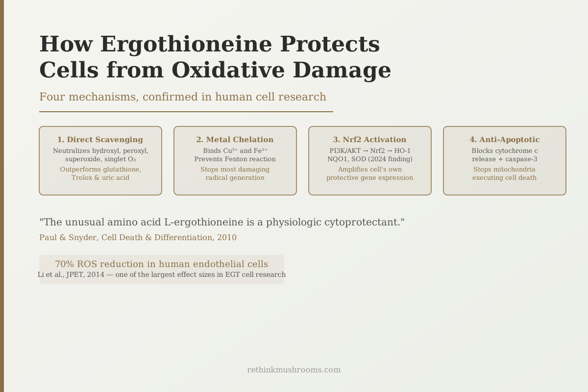
Ergothioneine reduced oxidative-stress-induced reactive oxygen species by up to 70% in human endothelial cells and prevented oxidative cell death, one of the largest effect sizes in ergothioneine cell biology research.
Li et al., Journal of Pharmacology and Experimental Therapeutics, 2014
4 mechanisms
direct scavenging, metal chelation, Nrf2 activation, anti-apoptotic, each confirmed in separate studies
>50%
reduction in pro-inflammatory cytokines TNF-α and IL-17 via macrophage modulation (Song et al., 2025)
2024
year the PI3K/AKT/Nrf2 signaling pathway was confirmed as ergothioneine's upstream mechanism in UV damage protection
Li et al., Journal of Pharmacology and Experimental Therapeutics, 2014
 View as image ↗
In 2010, Solomon Snyder and colleagues at Johns Hopkins published a paper in Cell Death and Differentiation with an unusually direct title: "The unusual amino acid L-ergothioneine is a physiologic cytoprotectant."[1] The framing was deliberate. Ergothioneine was not being described as a supplement or a nutraceutical with possible benefits; it was being named a compound the body uses as part of its native protection system. Cells actively accumulate it via OCTN1. When it is present, they survive toxic insults that would otherwise kill them.
This article covers the mechanisms behind that protection: how ergothioneine protects cells from oxidative damage, what chemistry makes it different from other antioxidants, what the most recent research adds, and where the honest limits of the evidence sit.
For background on what makes ergothioneine biologically unique, including the dedicated transporter OCTN1 that delivers it to high-stress tissues, see what ergothioneine is and why the body hoards it.
Key Takeaways
- Paul and Snyder (2010) in Cell Death and Differentiation named ergothioneine a "physiologic cytoprotectant": cells actively accumulate it via OCTN1 and use it to survive toxic oxidative insults
- Li et al. (2014) found ergothioneine reduced ROS by up to 70% in human endothelial cells, prevented oxidative cell death, and maintained endothelial nitric oxide synthase activity under pro-oxidant conditions
- Ergothioneine's thione sulfur form does not produce a depleted disulfide when it scavenges radicals, unlike glutathione, making its protection chemically self-renewing
- Nguyen et al. (2024) identified the PI3K/AKT/Nrf2 signaling pathway as the mechanism by which ergothioneine upregulates HO-1 and NQO1 antioxidant enzymes in UV-damaged skin cells
- Song et al. (2025) showed ergothioneine reduces inflammation through a non-antioxidant mechanism: shifting macrophages from pro-inflammatory M1 to anti-inflammatory M2 state, cutting TNF-α and IL-17 by more than 50%
- Most evidence comes from cell culture; the 2020 Singapore cardiovascular cohort is the strongest human correlate
Ergothioneine Is a Physiologic Cytoprotectant, Not Just an Antioxidant
Most antioxidant claims are made by supplement companies. The "physiologic cytoprotectant" label came from a laboratory that studies cell death mechanisms, published in a journal dedicated to that topic, based on direct experiments showing that cells without ergothioneine die more easily.
Paul and Snyder (2010) showed that OCTN1-expressing cells accumulated ergothioneine and used it to protect against DNA damage, oxidative stress, and cell death induced by multiple toxic agents including hydrogen peroxide (a model oxidative stressor) and mitomycin C (a DNA-damaging chemotherapy agent).[1] Cells that could not take up ergothioneine (because OCTN1 was blocked or absent) died at higher rates under the same conditions.
The study established three things that underpin the rest of the mechanism story: cells actively seek ergothioneine, they use it specifically against toxic threats, and without it they are more vulnerable. This is the strongest evidence that ergothioneine's protective effects are not incidental chemistry; they are part of how cells manage oxidative stress.
The Four Mechanisms: How Ergothioneine Protects Cells
Ergothioneine protects cells from oxidative damage through four distinct mechanisms, each operating through a different pathway:
- Direct free-radical scavenging: ergothioneine neutralizes hydroxyl radicals, peroxyl radicals, superoxide, singlet oxygen, and hypochlorous acid directly, faster than glutathione or Trolox (vitamin E analogue) at equivalent concentrations
- Metal chelation: ergothioneine binds pro-oxidant metal ions including copper (Cu²⁺) and iron (Fe²⁺), preventing them from catalyzing Fenton reactions that generate the most damaging reactive oxygen species
- Nrf2 pathway activation: ergothioneine activates Nrf2 (nuclear factor erythroid 2-related factor 2), a master transcription factor that upregulates the cell's own antioxidant enzyme production including HO-1, NQO1, catalase, and superoxide dismutase
- Anti-apoptotic protection: ergothioneine prevents cytochrome c release from mitochondria and caspase-3 activation, blocking the mitochondria-driven cell death pathway before it commits the cell to apoptosis
Cheah and colleagues' 2020 comprehensive biology review in Nutrition Research Reviews documented all four mechanisms with literature evidence.[5] Chen and colleagues' 2025 review in Food & Function integrated the most recent mechanistic findings including the Nrf2 pathway data.[6]
The Chemistry That Makes Ergothioneine's Protection Self-Renewing
Most antioxidants work by sacrificing themselves. Vitamin C gives up electrons and becomes dehydroascorbate. Glutathione scavenges radicals and forms glutathione disulfide (GSSG), a depleted form that must be enzymatically recycled before it can scavenge again. Every scavenging event depletes the available pool.
Ergothioneine is chemically different, and the difference starts at the sulfur atom. In solution at physiological pH, ergothioneine adopts the thione form, a different oxidation state of sulfur than the thiol (-SH) that glutathione uses. Servillo and colleagues (2017), in a Journal of Cardiovascular Pharmacology review, characterized this chemistry: ergothioneine scavenges free radicals without forming a stable oxidized disulfide product analogous to GSSG.[3]
In practical terms: ergothioneine's antioxidant capacity is not progressively consumed by each radical it neutralizes in the way that other common antioxidants are. This self-renewing chemistry is the basis for why Cheah and Halliwell found ergothioneine showed superior free radical scavenging activity compared to glutathione, Trolox (vitamin E analogue), and uric acid in head-to-head comparisons.[4]
The comparison to glutathione specifically is explored in depth in ergothioneine vs. glutathione: comparing master antioxidants, including the 85% vs. 20% bioavailability difference and the evidence for Nrf2-mediated synergy. For comparison with a different class of mushroom antioxidants, enoki phenolic compounds and oxidative stress protection covers how enoki's phenolic antioxidants work through a different chemistry than ergothioneine's thione mechanism.
In Endothelial Cells: The 70% ROS Reduction
Endothelial cells line the interior of blood vessels. Oxidative damage to these cells is an early step in cardiovascular disease, and ergothioneine's endothelial protection data is among the most direct human-cell evidence available.
Li and colleagues (2014) exposed human endothelial cells to pro-oxidant conditions, then measured the response with and without ergothioneine pretreatment.[2] Ergothioneine reduced reactive oxygen species by up to 70%, prevented oxidative cell death, and maintained endothelial nitric oxide synthase (eNOS) activity. eNOS produces nitric oxide, which regulates vascular tone and prevents clotting. Preserving eNOS activity under oxidative stress is a meaningful functional endpoint, not just a marker; it is what keeps blood vessels functioning normally.
In the same endothelial cell model, Servillo and colleagues (2017) showed ergothioneine traps superoxide radicals under diabetic (high-glucose) conditions, forming stable oxidation products far less reactive than the original radicals.[9] This safe neutralization, forming benign products rather than reactive intermediates, reduces secondary cellular damage that can occur when less stable radical reactions produce more harmful byproducts.
Aruoma and colleagues (2018) extended the endothelial protection evidence to toxic metal exposure: ergothioneine pretreatment fully prevented endothelial dysfunction caused by mercury chloride, demonstrating cytoprotection via both ROS scavenging and the metal chelation mechanism.[8] The Fenton reaction prevention (metal chelation) becomes most important in this scenario because toxic heavy metals like mercury drive massive free radical production through metal-catalyzed oxidation.
The metabolic disease context for this endothelial protection is covered in mushroom antioxidants and advanced glycation end products in diabetes, which places ergothioneine's high-glucose protection within the broader mushroom antioxidant story.
The 1983 Finding That Started the Mitochondrial Story
One of the earliest mechanistic observations about ergothioneine came from Aruoma and colleagues in 1983: ergothioneine inhibited lipid peroxide formation in isolated mitochondrial membranes more effectively than glutathione at equivalent concentrations.[7]
This 40-year-old finding is low-evidence by modern standards; the methods available in 1983 were limited. But it was the first observation that pointed ergothioneine's protective activity toward mitochondria specifically. Four decades of mechanistic work have since confirmed and extended that observation, culminating in the 2025 MPST identification (covered in ergothioneine and mitochondrial protection) and the 2025 cGPDH persulfidation pathway (covered in ergothioneine: preserving your cellular powerhouses).
Chen and colleagues (2021) captured the mitochondrial protection mechanism in action in endothelial cells: ergothioneine reduced mitochondrial ROS production, prevented cytochrome c release, and inhibited caspase-3 activation in cells challenged with toxic oxidized cholesterol.[10] These three events are sequential steps in the mitochondria-driven apoptosis pathway. Blocking all three early in the cascade is consistent with the cytoprotectant label Paul and Snyder applied: ergothioneine intercepts the death signal before it becomes committed.
Nrf2 Activation: The 2024 PI3K/AKT Upstream Pathway
The Nrf2 activation mechanism had been noted in several ergothioneine reviews, but Nguyen and colleagues (2024) were the first to trace the full signaling pathway upstream of Nrf2 in an ergothioneine study.[12] The context was UV-B damage in human skin keratinocytes.
UV-B radiation generates reactive oxygen species that damage keratinocyte DNA and proteins, causing cell death. Nguyen's team found that ergothioneine activated the PI3K/AKT signaling pathway, which in turn activated Nrf2. Nrf2 then upregulated two antioxidant enzymes: HO-1 (heme oxygenase-1) and NQO1 (NADPH quinone oxidoreductase 1). These enzymes are part of the cell's endogenous antioxidant enzyme system, not just direct scavengers but regulators that sustain long-term protective capacity.
The finding is important for two reasons. First, it identifies the exact molecular pathway from ergothioneine exposure to Nrf2 gene expression, which had not been mapped before 2024. Second, it confirms that ergothioneine's cellular protection extends far beyond direct radical scavenging: by activating PI3K/AKT/Nrf2, it switches on the cell's own protective machinery.
Cheah and Halliwell's 2022 Antioxidants and Redox Signaling paper noted that Nrf2 activation by ergothioneine also upregulates SIRT1 and SIRT6 expression.[11] SIRT1 and SIRT6 are sirtuins, proteins involved in DNA repair and aging-related gene regulation. This connects the direct cell protection mechanisms to the broader longevity biology discussed in the companion articles in this cluster.
For the full selectivity story linking OCTN1 to where ergothioneine goes in the body, see ergothioneine: the most selective antioxidant in your body.
Beyond Chemistry: The 2025 Immune Modulation Finding
For most of its research history, ergothioneine's cellular protection was understood through direct chemistry: it scavenges radicals, chelates metals, and activates antioxidant pathways. A 2025 paper by Song and colleagues added an entirely different dimension.
Song's team tested ergothioneine in a mouse model of psoriasis and found that it reduced psoriatic inflammation by shifting macrophage polarization from the pro-inflammatory M1 state to the anti-inflammatory M2 state.[13] Macrophages drive inflammatory skin disease through cytokine production. In the ergothioneine-treated animals, TNF-α and IL-17 (two key pro-inflammatory cytokines that drive psoriatic lesions) fell by more than 50%.
This is a cell-protection mechanism, but it operates through immune modulation rather than direct radical chemistry. Oxidative stress and chronic inflammation are linked processes: reactive oxygen species activate pro-inflammatory signaling, and inflammation generates more reactive oxygen species. By shifting macrophage state, ergothioneine appears to break this cycle indirectly.
Whether this M1/M2 effect is relevant beyond skin inflammation has not been studied. The psoriasis model is animal data, and the human relevance is unknown. But the finding expands ergothioneine's mechanism profile beyond what was known before 2025 and is consistent with the broader picture of a compound the body uses for active cellular defense.
What the Evidence Supports
The mechanism evidence for ergothioneine's cellular protection is unusually specific and well-documented for a dietary antioxidant. Paul and Snyder's "physiologic cytoprotectant" label holds up: cells accumulate ergothioneine via OCTN1 and use it against oxidative stress through at least four documented pathways. The chemistry is distinct (thione form, self-renewing), the cell data in human endothelial cells is concrete (70% ROS reduction, maintained eNOS), and the 2024 PI3K/AKT/Nrf2 work puts an upstream signaling pathway behind the Nrf2 activation that had previously been asserted rather than traced.
The honest limit: virtually all of this evidence is from cell culture (in vitro). There are no randomized controlled trials testing ergothioneine against a specific cellular oxidative damage endpoint in humans. The Singapore cardiovascular cohort provides the most compelling human correlate: 68% lower cardiovascular mortality in people with higher ergothioneine levels. But association studies cannot establish which mechanism, if any, drives that outcome.
The mechanism picture is strong. The translation to human clinical benefit is inferred, not yet demonstrated in trials.
The Bottom Line
Ergothioneine's cellular protection rests on four confirmed mechanisms: direct radical scavenging superior to glutathione and Trolox at equivalent concentrations, metal chelation blocking Fenton reaction-derived hydroxyl radicals, Nrf2/PI3K/AKT pathway activation upregulating the cell's own antioxidant enzymes, and anti-apoptotic action blocking cytochrome c release from mitochondria. The thione sulfur form makes its scavenging self-renewing, unlike glutathione's GSSG cycle. A 2025 paper added immune modulation through macrophage polarization as a fifth non-chemical mechanism. Human clinical trials with cellular oxidative damage endpoints do not yet exist; the 70% ROS reduction in human endothelial cells and the Singapore cardiovascular association are the strongest evidence that the mechanisms matter in practice.
Frequently Asked Questions
Sources & References
- 1. Source details for this reference are not yet available.
- 2. Source details for this reference are not yet available.
- 3. Source details for this reference are not yet available.
- 4. Source details for this reference are not yet available.
- 5. Source details for this reference are not yet available.
- 6. Source details for this reference are not yet available.
- 7. Source details for this reference are not yet available.
- 8. Source details for this reference are not yet available.
- 9. Source details for this reference are not yet available.
- 10. Source details for this reference are not yet available.
- 11. Source details for this reference are not yet available.
- 12. Source details for this reference are not yet available.
- 13. Source details for this reference are not yet available.