Maitake & Blood Sugar / Diabetes
3 distinct mechanisms
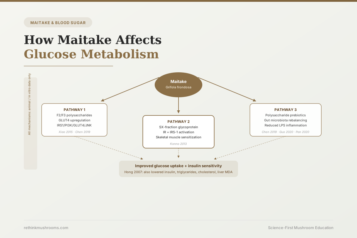
Maitake polysaccharides lower blood sugar through at least three mechanistically separate pathways that have been characterized across 30 years of research, and the gut microbiota pathway, documented only since 2019, is absent from virtually all consumer-facing content.
Chen et al., Food Chem Toxicol, 2019; Xiao et al., Food Funct, 2015; Konno et al., Int J Gen Med, 2013
GLUT4
glucose transporter upregulated by maitake F2/F3 polysaccharides in liver tissue (Xiao 2015)
IRS-1/Akt
insulin signaling proteins activated by SX-fraction glycoprotein in skeletal muscle (Konno 2013)
7 patients
the largest published human dataset on SX-fraction glycemic effects, a pilot study (Konno 2013)
Chen et al., Food Chem Toxicol, 2019; Xiao et al., Food Funct, 2015; Konno et al., Int J Gen Med, 2013
 View as image ↗
Most content about maitake and blood sugar says something vague about beta-glucans and insulin and stops there. The actual research is more specific and more interesting. Maitake (Grifola frondosa) contains at least three structurally different compounds that affect blood glucose through three mechanistically separate pathways. They work independently. They were discovered at different times by different research groups. And one of them, the gut microbiota pathway, was only characterized starting in 2019, which is why virtually no competitor content covers it.
This article is organized by mechanism, not by history. Each pathway gets its own section with the specific compounds, molecular targets, and experimental evidence. The evidence is almost entirely from animal models; that context appears honestly in each section. For the question of what clinical and human data exists, see Maitake and Blood Sugar: Research on Glycemic Control. For the foundational overview of what maitake is, see What Is Maitake Mushroom: The Dancing Mushroom Explained.
Key Takeaways
- Maitake affects glucose metabolism through at least three distinct pathways: (1) polysaccharide-mediated GLUT4 upregulation in liver and muscle, (2) glycoprotein-mediated insulin receptor sensitization via the IRS-1/Akt pathway, and (3) gut microbiota rebalancing that reduces inflammation-driven insulin resistance
- Xiao and colleagues (2015) directly measured increased GLUT4 expression in hepatic tissue of diabetic rats treated with maitake F2 and F3 polysaccharide fractions, alongside improved oral glucose tolerance and decreased insulin resistance index
- Konno and colleagues (2013) identified the SX-fraction glycoprotein as the compound activating the insulin receptor (IR) and its downstream substrate IRS-1 in skeletal muscle cells, a mechanism entirely distinct from beta-glucan polysaccharide action
- Chen and colleagues (2019) were the first to show maitake polysaccharides improve glucose metabolism through gut microbiota rebalancing: increased Bifidobacterium and Lactobacillus, decreased Clostridium, and downstream suppression of LPS-driven inflammation
- Hong and colleagues (2007) showed that maitake alpha-glucan improved not just blood glucose but also serum insulin, triglycerides, cholesterol, and liver oxidative stress markers simultaneously in diabetic mice, the most comprehensive metabolic picture in a single study
- The human evidence base consists of one 2-patient letter (Konno 2001) and one 7-patient pilot study (Konno 2013); neither is a randomized controlled trial, and no human RCT for maitake glucose effects has been published
Before Knowing How It Works: Three Decades of Animal Evidence
The story of maitake's glucose effects begins in 1994, when Kubo and colleagues identified the first polysaccharide fraction in the fruit body with measurable anti-diabetic activity in animal models.[1] That founding observation established that the glucose-lowering effect existed. It did not explain which fraction was responsible or how it worked.
Horio and Ohtsuru followed in 2001 with a controlled experiment showing that maitake extract improved oral glucose tolerance in streptozotocin-induced diabetic rats, the standard animal model for type 1 and late-stage type 2 diabetes research.[2] The animals' blood glucose response to an oral glucose challenge was meaningfully blunted compared to untreated diabetic controls.
These two papers established the phenomenon. The three pathway articles below explain the mechanism.
Pathway 1: How Maitake Polysaccharides Upregulate GLUT4
GLUT4 (glucose transporter type 4) is the protein responsible for pulling glucose out of the bloodstream and into cells. It sits inside the cell when insulin is absent, then moves to the cell surface when insulin signals arrive, where it opens a channel for glucose entry. In type 2 diabetes, GLUT4 expression and translocation are impaired, which is a central reason blood glucose stays elevated after meals.
Xiao and colleagues measured GLUT4 expression directly in 2015, using two specific polysaccharide fractions (F2 and F3) isolated from maitake fruit body.[3] In streptozotocin-induced diabetic rats, both fractions significantly reduced fasting blood glucose and improved the oral glucose tolerance test, the standard measure of how well the body handles a sugar load. The insulin resistance index fell. And critically, GLUT4 protein expression in hepatic (liver) tissue was significantly elevated.
The hepatic GLUT4 finding is worth attention. Liver cells do not typically rely on GLUT4 the way muscle cells do, but in metabolically stressed states, hepatic GLUT4 expression plays a role in post-meal glucose clearance. Increased hepatic GLUT4 expression means the liver is participating more actively in removing glucose from circulation after eating.
The compounds driving this effect are polysaccharides, the same category as the beta-glucans responsible for maitake's immune effects. But these are specific polysaccharide fractions (F2 and F3) with distinct structural characteristics from the D-fraction polysaccharides studied for immune function. The fractions are separable and mechanistically distinct.
Pathway 2: The SX-Fraction Glycoprotein and Insulin Receptor Sensitization
The second pathway involves a completely different type of compound. The SX-fraction is a glycoprotein, not a polysaccharide. Glycoproteins are proteins with sugar chains attached. The SX-fraction has a molecular weight of approximately 20,000 Da and was first characterized as a glucose-active compound by the Preuss research group at Georgetown University.
Konno and colleagues completed the most definitive mechanistic work in 2013, using L6 skeletal muscle cells to test the SX-fraction's molecular targets.[4] The SX-fraction activated two specific proteins in the insulin signaling cascade. The first was the insulin receptor (IR) itself, the cell-surface protein that binds insulin and triggers the downstream signaling cascade. The second was IRS-1 (insulin receptor substrate 1), the immediate downstream mediator that IR activates when insulin binds. SX-fraction treatment increased phosphorylation of both proteins in the muscle cells.
The practical meaning: the SX-fraction was making skeletal muscle cells more responsive to insulin by amplifying the signal at the receptor level, not by mimicking insulin or increasing insulin secretion. Skeletal muscle is the primary site of post-meal glucose disposal in humans; approximately 80% of glucose absorbed after a carbohydrate-containing meal ends up in skeletal muscle. Increasing the efficiency of insulin receptor signaling in muscle cells has direct consequences for blood glucose control.
The same paper included a pilot clinical observation: 7 type 2 diabetes patients already taking oral medications added SX-fraction to their regimen for 2 to 4 weeks and showed a 30 to 63% decline in fasting blood glucose measurements. This is not a randomized controlled trial. It lacks a control group, randomization, blinding, or adequate sample size. But it is the only published data showing a glycemic response to SX-fraction in humans with diabetes. The in vitro mechanism (IR/IRS-1 activation) and the human observation (glucose decline) point in the same direction.
For a full accounting of the SX-fraction evidence arc, from the Georgetown animal studies through Konno's mechanism work, see Maitake SX-Fraction and Insulin Sensitivity.
Pathway 3: The Gut Microbiota Connection
The third mechanism was not proposed until 2019. Before then, maitake's glucose effects were explained entirely by direct cellular pathways (GLUT4 upregulation, insulin receptor sensitization). Chen and colleagues introduced a different explanation: the gut microbiome is an intermediary, and maitake polysaccharides lower blood sugar partly by changing which bacteria dominate the gut.[5]
Their study tested a novel polysaccharide fraction (GFP) from Grifola frondosa in a type 2 diabetic mouse model. The glucose-lowering effect occurred via the IRS1/PI3K/GLUT4/JNK signaling pathway (consistent with Pathway 1) but was also accompanied by significant shifts in gut bacteria composition. Bifidobacterium and Lactobacillus populations increased. Clostridium populations decreased. This shift matters because harmful bacteria in the gut (including certain Clostridium species) produce lipopolysaccharide (LPS), an inflammatory molecule that triggers TLR4 receptors on gut cells and systemically elevates TNF-alpha and IL-6. Chronic low-grade LPS-driven inflammation is one of the mechanisms by which gut dysbiosis contributes to insulin resistance. Reducing the bacteria that produce LPS reduces the inflammatory load that makes insulin less effective.
Guo and colleagues replicated the gut microbiota finding in 2020 using a high-fat diet plus streptozotocin model, showing both hypoglycemic and hypolipidemic effects alongside gut microflora modulation.[6] Fasting blood glucose and triglycerides fell simultaneously, and the pattern of gut bacterial shifts was consistent with the Chen 2019 findings.
Pan and colleagues also confirmed in 2020 that a maitake extract containing both polysaccharides and organic acids normalized glycolipid metabolism in rats while reshaping gut microbiota composition.[7] Their results identified the gut-glucose axis as a consistent mechanistic feature, not an artifact of one research group.
This pathway is not unique to maitake; other dietary polysaccharides including those from other mushrooms and plant fibers produce similar prebiotic effects. But it offers a third distinct explanation for why maitake's glucose effects might be broader and more sustained than the direct cellular mechanisms alone could explain.
The Comprehensive Metabolic Picture: More Than Blood Sugar
Most research on maitake and blood sugar focuses on glucose as the primary endpoint. Hong and colleagues expanded the picture significantly in 2007 with an alpha-glucan fraction from maitake fruit body in KK-Ay mice, a genetic model of type 2 diabetes and obesity.[8]
The intervention produced improvements across five separate metabolic markers. Fasting plasma glucose fell significantly. Serum insulin fell, indicating improved insulin sensitivity rather than compensatory hyperinsulinemia. Serum triglycerides fell. Total cholesterol fell. Liver malondialdehyde, a marker of oxidative damage in liver tissue, fell.
This is a clinically relevant combination. Type 2 diabetes rarely presents as an isolated glucose problem. The metabolic syndrome picture includes elevated triglycerides, abnormal cholesterol, liver involvement, and hypertension alongside hyperglycemia. A single intervention reducing all five markers simultaneously in an animal model is a stronger result than one that only affects glucose. It suggests the maitake alpha-glucan is engaging a broad metabolic regulatory pathway rather than a single specific target.
The liver MDA finding connects to a separate but related question about maitake's hepatoprotective properties. For more on maitake's effects on liver function, see Maitake and Liver Health: Hepatoprotective Effects. The simultaneous blood pressure and glucose effects seen in the broader Preuss group research connect to Maitake and Blood Pressure: Antihypertensive Research.
The Human Evidence: Two Reports, Neither a Controlled Trial
The mechanistic pathways above are all grounded in animal and cell culture experiments. The question of whether these pathways produce measurable glucose effects in humans is answered by a much thinner evidence base.
The first human report was a 2001 letter to the editor in the Journal of Orthomolecular Medicine, written by Konno and colleagues. Two type 2 diabetes patients showed approximately 30% reductions in blood glucose with whole maitake supplementation. This single observation from two patients is the only published human evidence for whole-mushroom maitake on blood glucose. It is not an experiment. It has no control group. It has no blinding, no randomization, and no statistical analysis. It is reported because it exists, not because it constitutes evidence.
The second human report is the Konno 2013 pilot with 7 patients already discussed in the SX-fraction section. Seven patients, no control group, measured over 2 to 4 weeks, with patients already on oral diabetes medications. The 30 to 63% decline in fasting glucose is plausible given the animal evidence for SX-fraction, but cannot be interpreted in isolation.
A 2020 review by Lindequist and Haertel surveyed clinical trial evidence for medicinal mushrooms across all species in type 2 diabetes and concluded that rigorous human RCTs are largely absent, not just for maitake, but across the field. The animal evidence for multiple mechanisms is solid and consistent. The human evidence base is not there yet.
For the complete clinical evidence accounting and an honest evidence-tier assessment, see Maitake for Type 2 Diabetes: Clinical Evidence and Mechanisms. For the Georgetown research specifically and how it has been mischaracterized, see Maitake and Glucose Tolerance: The Georgetown University Clinical Research.
For broader pharmacological context on how Grifola frondosa works, see The Pharmacology of Grifola frondosa: How Maitake Works.
The Bottom Line
Maitake affects glucose metabolism through three mechanistically distinct pathways: (1) F2/F3 polysaccharides upregulate GLUT4 expression in liver and muscle tissue, improving cellular glucose uptake; (2) the SX-fraction glycoprotein activates the insulin receptor and IRS-1 in skeletal muscle, improving insulin sensitivity at the receptor level; (3) polysaccharides rebalance gut microbiota by promoting beneficial Bifidobacterium and Lactobacillus while reducing LPS-producing bacteria, thereby reducing inflammation-driven insulin resistance. Comprehensive animal studies show simultaneous improvements in glucose, insulin, triglycerides, cholesterol, and liver oxidative stress markers. The human evidence is limited to a 2-patient letter (Konno 2001) and a 7-patient uncontrolled pilot (Konno 2013); no randomized controlled trial has been published for maitake glucose effects in humans.
Frequently Asked Questions
Sources & References
- 1RTHM-00015·Kubo, K et al. (1994). “Anti-diabetic activity present in the fruit body of Grifola frondosa (Maitake). I..” Biological & pharmaceutical bulletin.Study breakdown →PubMed →↩
- 2RTHM-00035·Horio, H et al. (2001). “Maitake (Grifola frondosa) improve glucose tolerance of experimental diabetic rats..” Journal of nutritional science and vitaminology.Study breakdown →PubMed →↩
- 3RTHM-00410·Xiao, Chun et al. (2015). “Hypoglycemic effects of Grifola frondosa (Maitake) polysaccharides F2 and F3 through improvement of insulin resistance in diabetic rats..” Food & function.Study breakdown →PubMed →↩
- 4RTHM-00533·Konno, Sensuke et al. (2013). “Possible hypoglycemic action of SX-fraction targeting insulin signal transduction pathway..” International journal of general medicine.Study breakdown →PubMed →↩
- 5RTHM-01042·Chen, Yuqing et al. (2019). “Hypoglycemic activity and gut microbiota regulation of a novel polysaccharide from Grifola frondosa in type 2 diabetic mice..” Food and chemical toxicology : an international journal published for the British Industrial Biological Research Association.Study breakdown →PubMed →↩
- 6RTHM-01193·Guo, Wei-Ling et al. (2020). “Hypoglycemic and hypolipidemic activities of Grifola frondosa polysaccharides and their relationships with the modulation of intestinal microflora in diabetic mice induced by high-fat diet and streptozotocin..” International journal of biological macromolecules.Study breakdown →PubMed →↩
- 7RTHM-01272·Pan, Yuyang et al. (2020). “Regulatory effect of Grifola frondosa extract rich in polysaccharides and organic acids on glycolipid metabolism and gut microbiota in rats..” International journal of biological macromolecules.Study breakdown →PubMed →↩
- 8RTHM-00113·Hong, Lei et al. (2007). “Anti-diabetic effect of an alpha-glucan from fruit body of maitake (Grifola frondosa) on KK-Ay mice..” The Journal of pharmacy and pharmacology.Study breakdown →PubMed →↩