Meshima & Cancer
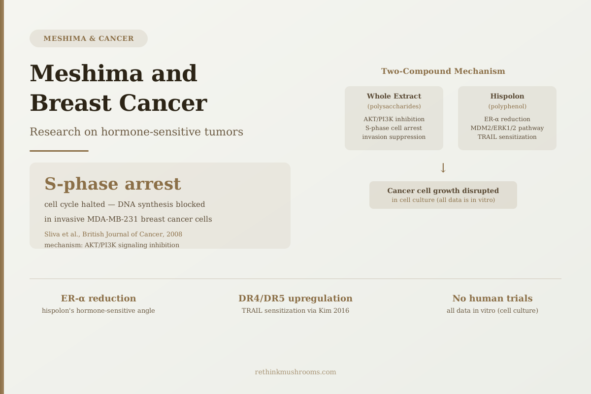
S-phase arrest
Sliva and colleagues showed Meshima extract halted highly invasive MDA-MB-231 breast cancer cell proliferation at the S-phase of the cell cycle — blocking DNA synthesis and suppressing the proteins cells use to invade surrounding tissue.
Sliva et al., British Journal of Cancer, 2008
AKT/PI3K inhibition
central anti-proliferative mechanism in breast cancer cells (Sliva 2008)
ER-α reduction
hispolon reduces estrogen receptor expression — relevant to hormone-sensitive tumors (Lu 2009)
TRAIL sensitization
hispolon upregulates DR4/DR5 death receptors, increasing immune killing (Kim 2016)
Sliva et al., British Journal of Cancer, 2008
 View as image ↗
Most of the mushroom-and-cancer research literature covers immune activation — beta-glucans stimulating NK cells and macrophages. Meshima (Phellinus linteus) has a different and more specific story in the breast cancer literature. The research describes two compound-level mechanisms that act directly on breast cancer cells: one through whole-extract polysaccharides that disrupt cell survival signaling, and one through hispolon, a polyphenol that reduces the expression of the estrogen receptor that drives hormone-sensitive tumors.
This article covers what the cell culture research on meshima breast cancer actually shows. The evidence is preclinical — entirely in vitro — and there are no human clinical trials of Meshima in breast cancer patients. That gap matters enormously, and the article holds to that boundary throughout. But the cell-level mechanisms documented across three independent research groups represent a more specific and interesting picture than competitor sites have presented.
For background on Meshima's general research history and why it's been studied for decades in Japan and Korea, see what Meshima mushroom is and why it's called Korea's anti-cancer mushroom.
Key Takeaways
- Sliva et al. (2008) tested Meshima extract against MDA-MB-231 cells — one of the most aggressive, invasive breast cancer cell lines — and found AKT/PI3K signaling inhibition with S-phase cell cycle arrest
- Invasion suppression was also documented: Meshima reduced the urokinase-plasminogen activator that MDA-MB-231 cells use to break through tissue barriers
- Hispolon, Meshima's key polyphenol, reduces estrogen receptor alpha (ER-α) expression through the MDM2/ERK1/2 pathway — a finding specifically relevant to hormone-sensitive (ER+) breast cancer
- A separate hispolon study (Kim 2016) showed it sensitizes cancer cells to TRAIL — an immune protein that naturally triggers cancer cell death — by upregulating the death receptors DR4 and DR5
- All breast cancer research for Meshima is in cell culture; no animal models of breast cancer specifically and no human trials have been published
- The AKT pathway Meshima inhibits is the same pathway targeted by some approved targeted therapies, which has implications for potential interactions
What Sliva's 2008 Study Found With the Most Aggressive Breast Cancer Cell Line
Not all breast cancer cell lines are equivalent in terms of what makes them useful for research. MDA-MB-231 cells, which Sliva and colleagues chose for their 2008 study, represent a particularly aggressive subtype: highly invasive, able to spread to secondary sites, and negative for estrogen receptors, progesterone receptors, and HER2 — making them one of the harder cancers to treat with targeted therapies.[1]
Working with this challenging cell line, the Sliva group showed that Phellinus linteus extract inhibited proliferation, reduced the formation of new cell colonies, and suppressed the invasive behavior that makes MDA-MB-231 cells clinically dangerous.
The central mechanism was inhibition of AKT/PI3K signaling. The AKT pathway is a survival signal that cancer cells exploit to keep dividing and resist programmed cell death. By interrupting this signal, Meshima extract prevented the cells from progressing through the cell cycle — specifically, they arrested at the S phase, the stage during which cells copy their DNA before dividing. The mechanism for that arrest was traced to upregulation of p27Kip1, a protein that acts as a brake on the cell cycle machinery.
Separately, the same study found that Meshima suppressed secretion of urokinase-plasminogen activator (uPA). uPA is an enzyme that cancer cells use to degrade the extracellular matrix — the structural scaffolding of tissue — making it possible for them to invade beyond the original tumor site. Reduced uPA activity is a proxy for reduced invasion potential. In Sliva's experiment, Meshima-treated MDA-MB-231 cells were significantly less able to invade through a membrane barrier than untreated cells.
This invasive behavior suppression is developed in more depth in Meshima and cancer metastasis: can it prevent spread?. For the full account of how Meshima inhibits tumor growth across cancer types, see Meshima and cancer: the complete research review.
Hispolon and Estrogen Receptors: The Hormone-Sensitive Angle
The finding that sets this article apart from any competitor coverage is the hispolon-estrogen receptor connection.
About 70% of breast cancers are estrogen receptor-positive (ER+). In these tumors, estrogen binds to estrogen receptor alpha (ER-α) and promotes cancer cell growth. Standard treatment for ER+ breast cancer includes drugs that either block estrogen production (aromatase inhibitors) or block the receptor itself (tamoxifen). The ER-α receptor is the direct therapeutic target.
Lu and colleagues found in 2009 that hispolon — a polyphenol compound found in Phellinus linteus — reduces ER-α expression through the MDM2/ERK1/2 pathway in breast cancer cells.[2] MDM2 is a protein that regulates p53, the tumor suppressor gene — but it also interacts with ER signaling. Hispolon's action via MDM2 recruitment changes the cellular environment in a way that reduces how much ER-α is expressed on the cancer cell surface.
The practical implication of reduced ER-α expression, if the mechanism held in living tissue, would be less estrogen-driven growth signaling in hormone-sensitive tumors. The cells would have fewer receptors to bind estrogen, reducing one of their core growth stimuli.
No competitor article has covered this angle. The Lu 2009 paper is occasionally cited in academic reviews for its MDM2/ERK1/2 mechanism, but the estrogen receptor relevance — the reason it matters specifically for ER+ breast cancer — is not translated for lay readers anywhere else in publicly available consumer content.
One more nuance from Lu's study: hispolon's MDM2 action also explains why it can trigger apoptosis in cells with altered or mutated p53. Many cancer cells develop mutations in p53 that reduce normal cell death signals. Hispolon works through MDM2 rather than requiring functional p53, which gives it potential activity across a broader range of cancer cell types than compounds that depend on the p53 pathway being intact.
How Hispolon Makes Cancer Cells More Vulnerable to Immune Killing
A third mechanism came from Kim and colleagues in 2016, testing hispolon against multiple cancer cell lines in Oncology Reports.[3]
TRAIL — tumor necrosis factor-related apoptosis-inducing ligand — is a protein produced by the immune system that induces apoptosis in cancer cells. Immune cells, particularly NK cells and T cells, use TRAIL as a weapon against tumor cells. One reason some cancers evade immune killing is that they downregulate the death receptors that TRAIL binds to — DR4 and DR5.
Kim's study found that hispolon upregulates DR4 and DR5 in cancer cells. With more death receptors on the surface, the cells become substantially more susceptible to TRAIL-induced cell death. In practical terms: hispolon may restore a killing pathway that some cancer cells have learned to evade.
This finding connects the direct chemical activity of hispolon to the immune-mediated anti-cancer mechanisms that the broader Meshima research covers. The polysaccharide components of Meshima activate immune cells including NK cells, which then use TRAIL. The hispolon component appears to make cancer cells more vulnerable to what those NK cells are delivering.
For details on how Meshima triggers apoptosis through caspase pathways, including the TRAIL connection, see Meshima and cancer cell apoptosis: how it triggers cell death.
Two Compounds, Two Pathways: Why the Distinction Matters
It's worth making explicit what the research establishes about these two branches of Meshima's breast cancer activity, because they have different implications.
The whole-extract Meshima research (Sliva 2008) works through polysaccharide-mediated disruption of AKT/PI3K signaling. This is the pathway most directly compared to targeted therapy drugs like PI3K inhibitors (alpelisib/Piqray) approved specifically for ER+/HER2- breast cancer.
The hispolon research (Lu 2009, Kim 2016) works through a different set of pathways — MDM2/ERK1/2 for ER-α reduction, and direct caspase activation and death receptor upregulation for apoptosis. These are chemically specific to the polyphenol compound rather than the polysaccharide fraction.
Sliva's comprehensive 2010 review of Phellinus linteus research noted that it has shown the strongest antitumor activity among mushrooms tested in 40 years of Japanese research — and that this activity appears through multiple compound classes and multiple mechanisms rather than a single dominant pathway.[4]
The distinction between polysaccharide and hispolon mechanisms matters for product selection. A whole-extract supplement preserves both compound classes. Products that fractionate extracts to isolate only polysaccharides or only specific biomarkers may lose the hispolon content that drives the ER-α reduction and TRAIL sensitization findings.
The In Vivo Evidence Gap — and What the Closest Animal Data Shows
The honest situation with Meshima and breast cancer is that the laboratory data stops at cell culture. No published animal model study has tested Meshima specifically against breast cancer tumors in vivo. The only in vivo Meshima cancer data in the literature involves prostate cancer xenografts and hepatoma models.
Tsuji and colleagues demonstrated in 2010 that Phellinus linteus extract caused tumor regression in athymic nude mice bearing advanced prostate cancer xenografts, and sensitized those tumors to doxorubicin-induced cell death through caspase-mediated pathways.[5] This is the most rigorous in vivo anti-tumor evidence for Meshima in any cancer type — not breast cancer, but demonstrating that the cell-culture mechanisms translate to measurable tumor activity in a living animal model.
Wang and colleagues (2023) worked with Sanghuangporus sanghuang, the taxonomic relative most closely related to Phellinus linteus, and showed inhibited proliferation and invasion of lung cancer cells in both cell culture and a mouse xenograft model.[6] This is the most recent published in vivo evidence from the Phellinus/Sanghuangporus species complex, and it demonstrates that the in vitro mechanisms do produce anti-tumor effects in at least one animal model context.
Neither of these studies is breast cancer data. Extrapolating from prostate or lung cancer xenografts to hormone-sensitive breast cancer involves assumptions about mechanism transfer that have not been tested. The cell culture work on breast cancer cells is internally consistent — three independent research groups found anti-tumor activity. The translation to animal models and then to human patients represents substantial additional steps that haven't been taken.
What the Evidence Gap Means for Anyone Researching Meshima and Breast Cancer
No randomized clinical trial of Meshima in breast cancer patients has been published. No observational study has tracked outcomes in breast cancer patients taking Meshima. The totality of breast cancer-specific evidence is three in vitro experiments, all showing interesting mechanisms, none establishing clinical efficacy.
This is worth stating plainly because the search queries that lead to this article come largely from people making real medical decisions. The cell culture evidence that Meshima inhibits AKT signaling, reduces ER-α expression, and sensitizes cells to TRAIL killing is scientifically interesting and mechanistically specific. It is not evidence that taking Meshima supplements produces these effects in breast cancer patients.
One additional consideration specific to hormone-sensitive breast cancer: the AKT/PI3K pathway that Sliva's 2008 study showed Meshima inhibiting is the same pathway targeted by approved PI3K inhibitors used in ER+/HER2- breast cancer treatment. Whether Meshima supplements could interact with these drugs — additively, synergistically, or antagonistically — has not been studied. This is exactly the kind of question that requires discussion with an oncologist.
For the broader evidence base on how Meshima works across cancer types, see Phellinus linteus and tumor growth inhibition: mechanisms. For readers comparing mushrooms with cancer research across multiple species, see how Turkey Tail enhances the immune system's anti-cancer response, which has a more developed human clinical evidence base.
The Bottom Line
Three independent research groups have documented anti-breast cancer activity from Meshima compounds in cell culture: Sliva 2008 showed AKT/PI3K inhibition and invasion suppression in MDA-MB-231 cells; Lu 2009 found hispolon reduces estrogen receptor alpha expression through MDM2/ERK1/2 — the hormone-sensitive breast cancer connection no competitor covers; Kim 2016 showed hispolon sensitizes cancer cells to TRAIL killing. All data is in vitro. No animal breast cancer model and no human clinical trial has been published. The mechanisms are specific and internally consistent, but the gap between cell culture and clinical evidence is large and has not been bridged.
Frequently Asked Questions
Sources & References
- 1RTHM-00271·Sliva, D et al. (2008). “Phellinus linteus Extract in Breast-Cancer Cell Models: What This 2008 Study Actually Found.” British journal of cancer.Study breakdown →PubMed →↩
- 2RTHM-00306·Lu, Te-Ling et al. (2009). “Hispolon from Phellinus linteus has antiproliferative effects via MDM2-recruited ERK1/2 activity in breast and bladder cancer cells..” Food and chemical toxicology : an international journal published for the British Industrial Biological Research Association.Study breakdown →PubMed →↩
- 3RTHM-00792·Kim, Ji-Hun et al. (2016). “Hispolon from Phellinus linteus induces apoptosis and sensitizes human cancer cells to the tumor necrosis factor-related apoptosis-inducing ligand through upregulation of death receptors..” Oncology reports.Study breakdown →PubMed →↩
- 4RTHM-00364·Sliva, Daniel (2010). “Medicinal mushroom Phellinus linteus as an alternative cancer therapy..” Experimental and therapeutic medicine.Study breakdown →PubMed →↩
- 5RTHM-00367·Tsuji, Takanori et al. (2010). “Phellinus linteus extract sensitizes advanced prostate cancer cells to apoptosis in athymic nude mice..” PloS one.Study breakdown →PubMed →↩
- 6RTHM-01817·Wang, Weike et al. (2023). “Sanghuangporus sanghuang extract inhibits the proliferation and invasion of lung cancer cells in vitro and in vivo..” Nutrition research and practice.Study breakdown →PubMed →↩