Meshima & Cancer
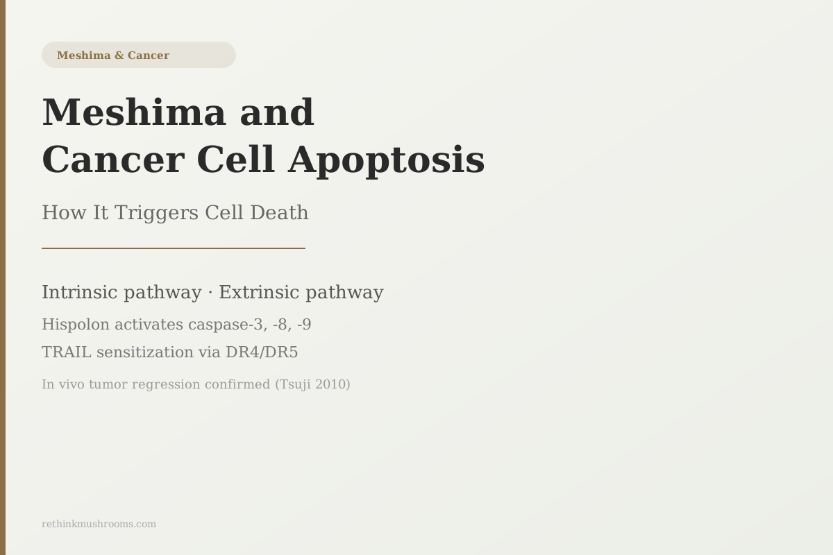
2 apoptosis pathways
In athymic nude mice bearing prostate cancer xenografts, Phellinus linteus injection both prevented new tumor formation and caused existing tumors to regress through caspase-mediated apoptosis.
Tsuji et al., PLoS One, 2010
3 caspases
caspase-3, -8, -9 activated by hispolon across cancer types (Chen 2013, Kim 2016)
DR4 + DR5
death receptors upregulated by hispolon, sensitizing cells to TRAIL (Kim 2016)
G0/G1
cell cycle arrest phase induced by hispolon before apoptosis (Chen 2013)
Tsuji et al., PLoS One, 2010
 View as image ↗
Cancer cells resist death. Normal cells carry built-in death programs that activate when the cell is damaged, infected, or no longer needed. Cancer cells mutate those programs, downregulate the proteins that trigger them, and develop resistance to the signals that would normally tell a compromised cell to self-destruct. The result is uncontrolled survival.
Two studies of Phellinus linteus, published in the British Journal of Cancer and PLoS One, identified specific mechanisms by which meshima's compounds bypass these resistances. One found that the extract activated different apoptosis routes depending on the cancer's hormone status. The other found that tumors in mice not only stopped forming but actually regressed when treated. These are the details the supplement industry glosses over and the academic literature buries in technical language.
This article covers what meshima apoptosis research actually shows, compound by compound, pathway by pathway. For background on meshima's other anti-cancer mechanisms, see Meshima and Anti-Angiogenesis. For a foundational introduction to the mushroom, see What Is Meshima Mushroom.
Key Takeaways
- Meshima compounds trigger cancer cell apoptosis through two distinct routes: the intrinsic pathway (driven by internal cellular damage, via mitochondria) and the extrinsic pathway (driven by external death signals, via surface receptors)
- Zhu et al. (2007, British Journal of Cancer) found Phellinus linteus activated different apoptotic mechanisms in prostate cancer cells depending on androgen receptor status: caspase-8 and ER stress in hormone-driven cells, a distinct partial pathway in castration-resistant cells
- Hispolon, Meshima's primary polyphenol, induced G0/G1 cell cycle arrest and apoptosis in leukemia cells via caspase-3 and caspase-9, with greater toxicity to cancer cells than to normal cells (Chen 2013)
- Kim et al. (2016) found hispolon sensitized multiple cancer cell lines to TRAIL, an immune protein that kills cancer cells, by upregulating death receptors DR4 and DR5 on the cancer cell surface
- Tsuji et al. (2010) confirmed in vivo tumor regression in athymic nude mice bearing advanced prostate cancer: PL injection attenuated new tumor growth and caused existing tumors to regress through caspase-mediated apoptosis
- All apoptosis data for Phellinus linteus is from cell culture or animal models; no human clinical trial has measured its effect on cancer cell apoptosis in patients
What Apoptosis Is and Why Cancer Cells Work to Defeat It
Apoptosis is programmed cell death: an ordered, controlled process by which the body removes cells that are damaged, infected, or surplus to requirements. It is not the same as necrosis, which is uncontrolled cell death from injury. Apoptosis is a built-in suicide program that healthy cells run when certain conditions are met.
Two separate pathways can trigger it. The intrinsic pathway is activated by signals from inside the cell: DNA damage, oxidative stress, or signals from the mitochondria (the cell's energy-generating organelles) that the cell is compromised beyond recovery. The extrinsic pathway is activated by signals from outside: binding of specific proteins to death receptors on the cell surface, which directly trigger the apoptosis cascade.
Both pathways converge on a family of proteins called caspases, which act as molecular scissors that dismantle the cell from the inside. Caspase-9 is typically activated by the intrinsic pathway; caspase-8 by the extrinsic pathway; caspase-3 is the final common executor that both activate. When caspase-3 is active, the cell breaks apart in an orderly way.
Cancer cells circumvent this system at multiple points. They overproduce proteins that block caspase activation, downregulate the death receptors on their surfaces, and suppress the mitochondrial signals that would trigger the intrinsic pathway. One of the core goals of cancer research is finding compounds that can reactivate apoptosis despite these defenses. A 2010 review by Sliva placed Phellinus linteus among the most studied mushrooms for this purpose, with antitumor activity documented across 40 years of Japanese and Korean research in prostate, breast, colon, lung, and melanoma models.[8]
The Prostate Cancer Studies: Why Androgen Receptor Status Changes the Response
The most specific published apoptosis data for Phellinus linteus comes from Zhu et al. (2007) in the British Journal of Cancer.[1] The study used two prostate cancer cell lines that represent clinically important subtypes: LNCaP cells, which are androgen receptor positive (meaning they depend on male hormones to grow), and PC3 cells, which are androgen receptor negative (meaning they have lost that hormone dependence and are typically more aggressive and drug-resistant).
The results differed between the two cell lines in a way that has clinical implications. In LNCaP cells, Phellinus linteus activated caspase-8 and a signaling partner called BID, which connects the extrinsic and intrinsic pathways. It also activated endoplasmic reticulum (ER) stress signaling. The ER is the cellular factory that folds proteins; when it's under stress, it can trigger apoptosis independently. In the hormone-dependent cells, PL was reaching apoptosis through two separate signals at once.
In PC3 cells (androgen receptor negative), PL still activated apoptosis, but through a different and less complete set of signals. The response was driven partly through caspase-2, which is activated downstream of the androgen receptor pathway. The finding: when the androgen receptor pathway is absent, PL's apoptotic reach into the cell is different.
Why this matters clinically: hormone-driven (androgen receptor positive) prostate cancer and castration-resistant (AR negative) prostate cancer are managed with entirely different treatment approaches. If a compound activates substantially different apoptotic pathways depending on hormone receptor status, the cancer type affects the expected response. This is a level of mechanistic nuance that consumer content uniformly ignores.
Hispolon: The Polyphenol Behind Multiple Apoptosis Pathways
Hispolon is a polyphenol found in Phellinus linteus. Unlike the polysaccharide fractions that dominate mushroom immune research, hispolon is a small molecule with a structure that allows it to interact with protein targets inside cells. It is the most studied individual compound from PL for anti-cancer activity.
Lu et al. (2009) characterized hispolon's mechanism in breast and bladder cancer cells using the MDM2/ERK1/2 pathway.[2] MDM2 is a protein that acts as a natural brake on p53, the tumor suppressor that would normally trigger apoptosis in response to cellular damage. When MDM2 is overexpressed (as it is in many cancers), it prevents p53 from functioning. Hispolon appears to recruit MDM2 through ERK1/2 signaling in a way that shifts the balance back toward apoptosis. The mechanistic significance: hispolon can push cancer cells toward death even when their p53 pathway is partially compromised.
Chen et al. (2013) studied hispolon in NB4 leukemia cells, a human acute promyelocytic leukemia line.[3] Hispolon induced G0/G1 cell cycle arrest (a hold at the cell's resting phase, preventing it from dividing) followed by apoptosis through the mitochondrial pathway, activating both caspase-3 and caspase-9. The study also compared hispolon's toxicity to cancer cells versus normal cells and found a meaningful selectivity: the compound was more toxic to NB4 leukemia cells than to normal control cells. This selectivity claim is made broadly in supplement marketing but rarely supported with data; the Chen 2013 study provides it.
Chen 2013 also showed that hispolon affected multiple intracellular targets, including the Bcl-2 family of proteins that normally inhibit mitochondrial apoptosis. By reducing Bcl-2 and related proteins, hispolon removed a key survival shield.
TRAIL Sensitization: Recruiting the Immune System
TRAIL (tumor necrosis factor-related apoptosis-inducing ligand) is a protein produced naturally by immune cells, particularly NK cells and cytotoxic T-cells. It kills cancer cells by binding to specific death receptors on their surface (DR4 and DR5) and triggering the extrinsic apoptosis pathway. The problem: many cancer cells downregulate DR4 and DR5, making themselves invisible to TRAIL-mediated killing. This is one mechanism cancer cells use to evade immune detection.
Kim et al. (2016) found that hispolon reversed this resistance in multiple cancer cell lines.[4] Treatment with hispolon upregulated DR4 and DR5 on cancer cell surfaces, making the cells significantly more susceptible to TRAIL-induced apoptosis. The combination of hispolon plus TRAIL killed more cancer cells than either treatment alone.
The implication is that hispolon may work not just as a direct apoptosis inducer, but also by making cancer cells more visible to the immune system's own cancer-killing mechanisms. A cell that downregulates its TRAIL receptors to hide from immune surveillance becomes, after hispolon treatment, a cell that is re-exposed. This is a distinct mechanism from everything discussed above and represents a potential combination approach with TRAIL-based therapies being explored in oncology.
For context on how this immune interaction connects to Meshima's broader immunomodulatory effects, see Meshima and Cancer Metastasis.
Two Apoptosis Routes, One Executioner
How Meshima’s Compounds Reach Caspase-3 From Two Directions
Cancer cells resist death by blocking apoptosis at several points. In lab studies, hispolon attacks from both sides at once: it tears down the mitochondrial survival shield inside the cell (the intrinsic route) and re-exposes the death receptors that let immune signals kill it from outside (the extrinsic route). Both lanes converge on caspase-3, the final executioner. Reading them side by side shows why one small molecule can push a resistant cell toward death by more than one mechanism.
Hispolon dismantles the cell’s survival shield from inside
Hispolon, meshima’s primary polyphenol, first holds leukemia cells at a G0/G1 arrest, then lowers the Bcl-2 family proteins that normally guard the mitochondria. With the shield down, the mitochondrial pathway fires caspase-9, which activates the executioner caspase-3. A related compound axis (MDM2/ERK1/2) lets hispolon push cells toward death even when their p53 brake is partly disabled.
Chen 2013 [3] (NB4 leukemia; caspase-9 + caspase-3; more toxic to cancer than normal cells) · Lu 2009 [2] (MDM2/ERK1/2, p53 axis) — all in vitro
Hispolon re-exposes cancer cells to the immune system’s kill signal
Many cancer cells hide from TRAIL — an NK/T-cell apoptosis signal — by downregulating its landing pads, the death receptors DR4 and DR5. Hispolon reverses that, pushing DR4/DR5 back onto the surface so TRAIL can bind, trigger caspase-8, and feed the same caspase-3 executioner. Hispolon plus TRAIL killed more cells than either alone.
Kim 2016 [4] (multiple cancer lines; DR4/DR5 up, caspase-8, TRAIL sensitization) — in vitro
Every molecular step shown is established only in cell culture or animal models. No human trial has measured apoptotic outcomes from Phellinus linteus, and the concentrations used in these studies are not directly translatable to oral doses in people. This map explains a documented laboratory mechanism — it is not evidence that meshima treats cancer in humans.
Mechanism synthesis; arms from refs [1][2][3][4][6] (see references)
The Polysaccharide Route: A Different Compound, a Different Cell Cycle Target
The studies above focus primarily on hispolon. But Phellinus linteus also contains protein-bound polysaccharides, and these take a different approach to inducing cancer cell death.
Li et al. (2004) tested a protein-bound polysaccharide fraction from PL on SW480 human colon cancer cells and found it induced G2/M phase cell cycle arrest followed by apoptosis.[5] The G2/M checkpoint is the one cells pass through just before dividing. Arresting here means the cell's DNA damage is detected at the last possible moment before copying, giving the apoptotic machinery one more chance to intervene.
The contrast with hispolon is notable. Hispolon (Chen 2013) arrests cells at G0/G1, the phase at the beginning of the cell cycle, before DNA replication starts. The polysaccharide arrests at G2/M, near the end. Both ultimately result in apoptosis, but through cell cycle checkpoints that represent entirely different points in the cellular process. The same mushroom uses two distinct timing strategies to stop cancer cell division.
Huang et al. (2011) added an important practical point: oral administration of PL mycelial culture produced antitumor effects in hepatoma-bearing mice.[7] Most cell culture and injection studies test compounds delivered directly to cells or injected into animals. An oral administration study validates that the compounds survive digestion and absorption and still produce measurable effects in a living organism. It is the delivery method closest to how humans actually consume the mushroom.
The In Vivo Evidence and What's Still Missing
Tsuji et al. (2010) provided the strongest available preclinical evidence for meshima's apoptotic anti-tumor activity.[6] In athymic nude mice bearing advanced prostate cancer xenografts, PL extract was injected intratumorally. Two outcomes were observed: new tumor formation was dramatically attenuated, and tumors that had already formed underwent regression via apoptosis, confirmed by caspase activation analysis. The study also found that PL sensitized the cancer cells to doxorubicin, a standard chemotherapy drug, with the combination producing more cell death than doxorubicin alone.
Tsuji et al. (2010) · PLoS One
- Population
- Athymic nude mice bearing advanced human prostate cancer xenografts
- Intervention
- Phellinus linteus extract injected directly into the tumor (intratumoral)
- Comparator
- Untreated tumors; and doxorubicin alone vs. doxorubicin + PL
- Finding
- New tumor formation was attenuated and existing tumors regressed via caspase-mediated apoptosis. PL also sensitized the cancer cells to doxorubicin, so the combination killed more cells than the chemotherapy drug alone.
preliminary Animal model with intratumoral injection — not an oral dose and not a human result. It shows the mechanism operates in a living organism, not that meshima treats cancer in people.
Tsuji et al., PLoS One (2010)
The sensitization finding echoes the TRAIL sensitization from Kim 2016 and suggests a pattern: PL compounds may broadly enhance cancer cells' susceptibility to death signals, both from the immune system and from chemotherapy. This is a potentially important clinical angle that remains unexplored in human trials.
Two honest gaps frame everything above:
No human apoptosis data exists. Every caspase activation result, every cell cycle arrest measurement, every TRAIL sensitization finding comes from cells growing in culture or from mice. The concentrations used in these studies are not directly translatable to oral doses in humans. Pharmacokinetics determine how much of any compound reaches a tumor in a living person after oral consumption, and those parameters have not been measured for PL's apoptosis-active fractions.
The 2024 hepatocellular carcinoma nuance. Chen et al. (2024) found that Phellinus linteus activated regulatory T cells (Treg) via FAK signaling and promoted M2 macrophage polarization in a hepatocellular carcinoma model, which could support tumor immune evasion rather than tumor destruction.[9] This finding applies specifically to hepatocellular carcinoma context and does not negate the apoptosis data in other cancer types, but it is an important signal that PL's effects are not uniformly anti-tumor across all cancer contexts. The complete cancer research picture, including this finding, is covered in Meshima and Cancer: The Complete Research Review.
For the broader comparison of Meshima against other cancer-relevant mushrooms with stronger clinical evidence, see How Turkey Tail Enhances the Immune System's Anti-Cancer Response.
Before you act on this
CriticalPreclinical apoptosis data is not evidence that meshima treats cancer in people
Concern
Every finding on this page comes from cancer cells in a dish or tumors in mice, often using injected extract at concentrations no oral supplement is known to reach in a human tumor. Some Phellinus linteus effects are context-dependent — in one liver-cancer model the mushroom appeared to help tumors evade the immune system rather than destroy them (Chen 2024). None of this has been tested as a cancer treatment in a human trial.
What the research says
Treat meshima as a research topic, not a therapy. If you have cancer, decisions about supplements belong in a conversation with your oncology team — not least because compounds that alter apoptosis and drug sensitivity can interact with chemotherapy and radiation.
Particularly relevant for: Anyone with a cancer diagnosis, in active treatment, or considering meshima alongside conventional care
What to do
Do not delay, replace, or modify any prescribed cancer treatment based on this article. Tell your oncologist before starting any mushroom supplement.
This is general educational information, not medical advice.
The Bottom Line
Phellinus linteus (meshima) triggers cancer cell apoptosis through multiple documented pathways: hispolon activates caspase-3, -8, and -9, induces G0/G1 arrest, and sensitizes cancer cells to immune-mediated TRAIL killing; protein-bound polysaccharides produce G2/M arrest via a distinct checkpoint; in vivo tumor regression has been confirmed in prostate cancer mouse models. The androgen receptor status of prostate cancer cells determines which apoptotic pathway PL activates. All evidence is preclinical; no human trial has measured apoptotic outcomes from Phellinus linteus.
Frequently Asked Questions
Sources & References
- 1RTHM-00240·Zhu, T et al. (2007). “Phellinus linteus activates different pathways to induce apoptosis in prostate cancer cells..” British journal of cancer.Study breakdown →PubMed →↩
- 2RTHM-00306·Lu, Te-Ling et al. (2009). “Hispolon from Phellinus linteus has antiproliferative effects via MDM2-recruited ERK1/2 activity in breast and bladder cancer cells..” Food and chemical toxicology : an international journal published for the British Industrial Biological Research Association.Study breakdown →PubMed →↩
- 3RTHM-00514·Chen, Yi-Chuan et al. (2013). “Hispolon from Phellinus linteus induces G0/G1 cell cycle arrest and apoptosis in NB4 human leukaemia cells..” The American journal of Chinese medicine.Study breakdown →PubMed →↩
- 4RTHM-00792·Kim, Ji-Hun et al. (2016). “Hispolon from Phellinus linteus induces apoptosis and sensitizes human cancer cells to the tumor necrosis factor-related apoptosis-inducing ligand through upregulation of death receptors..” Oncology reports.Study breakdown →PubMed →↩
- 5RTHM-00140·Li, Ge et al. (2004). “Protein-bound polysaccharide from Phellinus linteus induces G2/M phase arrest and apoptosis in SW480 human colon cancer cells..” Cancer letters.Study breakdown →PubMed →↩
- 6RTHM-00367·Tsuji, Takanori et al. (2010). “Phellinus linteus extract sensitizes advanced prostate cancer cells to apoptosis in athymic nude mice..” PloS one.Study breakdown →PubMed →↩
- 7RTHM-00403·Huang, Hui-Yu et al. (2011). “Orally administered mycelial culture of Phellinus linteus exhibits antitumor effects in hepatoma cell-bearing mice..” Journal of ethnopharmacology.Study breakdown →PubMed →↩
- 8RTHM-00364·Sliva, Daniel (2010). “Medicinal mushroom Phellinus linteus as an alternative cancer therapy..” Experimental and therapeutic medicine.Study breakdown →PubMed →↩
- 9RTHM-01867·Chen, Feihua et al. (2024). “Phellinus linteus activates Treg cells via FAK to promote M2 macrophage polarization in hepatocellular carcinoma..” Cancer immunology.Study breakdown →PubMed →↩