Meshima & Anti-Inflammatory Mechanisms
HO-1 induction
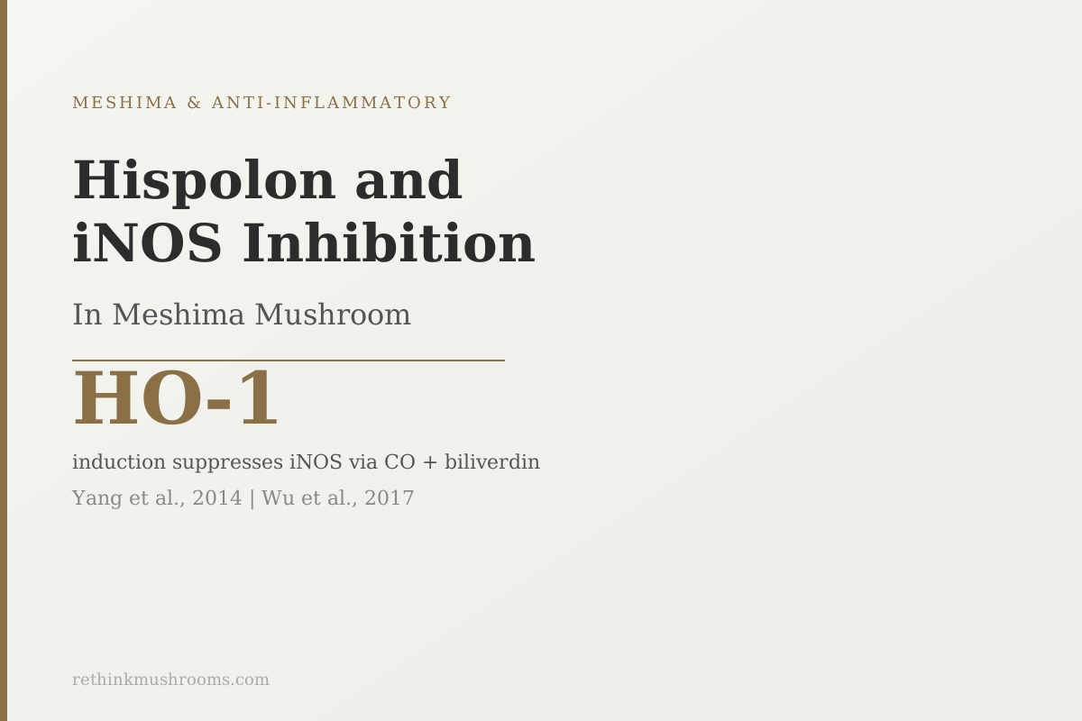
Hispolon doesn't block iNOS directly. It triggers HO-1, a protective enzyme whose byproducts (carbon monoxide and biliverdin) secondarily suppress inflammatory nitric oxide production.
Yang et al., Journal of Ethnopharmacology, 2014
iNOS/NO
suppressed in macrophages by hispolon via HO-1 (Yang 2014)
Brain
microglial cells respond to hispolon the same way as peripheral macrophages (Wu 2017)
2 pathways
hispolon (HO-1) and inotilone (MAPK/IKK) both inhibit iNOS in Meshima
Yang et al., Journal of Ethnopharmacology, 2014
 View as image ↗
Most anti-inflammatory compounds work by blocking the enzyme or signal that causes the problem. Hispolon, a phenolic compound isolated from Meshima (Phellinus linteus), takes an indirect route: instead of blocking iNOS (the enzyme that generates inflammatory nitric oxide) directly, hispolon triggers a protective enzyme called HO-1 whose natural byproducts are anti-inflammatory. This hispolon iNOS inhibition mechanism was first documented in macrophage studies and later confirmed in brain microglial cells, extending the finding to neuroinflammation: territory no consumer content on this mushroom has explored. This is a cluster article within the Meshima mushroom guide; all evidence reviewed here is preclinical, from cell and animal studies.
Key Takeaways
- iNOS (inducible nitric oxide synthase) is the enzyme activated during infection and chronic inflammation that produces damaging concentrations of nitric oxide (NO); blocking it is a recognized anti-inflammatory strategy
- Hispolon from Meshima suppresses iNOS-driven NO production and inflammatory cell death in macrophages by upregulating HO-1 (heme oxygenase-1), a protective enzyme (Yang et al., 2014)
- HO-1's products, carbon monoxide and biliverdin/bilirubin, carry the actual anti-inflammatory signal; hispolon induces this pathway rather than directly blocking iNOS
- In brain microglial cells (the immune cells of the brain), hispolon produced the same iNOS suppression and HO-1 upregulation, extending the mechanism to neuroinflammation (Wu et al., 2017)
- A second Meshima compound, inotilone, also inhibits iNOS through a different pathway: MAPK/IKK phosphorylation blocking, not HO-1 induction
- All findings are from cell cultures; no human trial has measured hispolon's effect on iNOS or nitric oxide levels in living people
What iNOS Is and Why Blocking It Reduces Inflammatory Damage
The body produces nitric oxide through a family of enzymes called nitric oxide synthases (NOS). Two of them, eNOS and nNOS, operate continuously at low levels and serve normal physiological functions: regulating blood vessel tone, acting as a neurotransmitter, and coordinating vascular responses to exercise.
The third, iNOS (inducible nitric oxide synthase), is different. It is not expressed under normal conditions. It turns on specifically in response to inflammatory signals, particularly lipopolysaccharide (LPS) from bacterial cell walls and pro-inflammatory cytokines. When activated, iNOS produces nitric oxide in quantities far exceeding what eNOS generates, creating concentrations that are damaging rather than regulatory.
At these high concentrations, NO reacts with superoxide to form peroxynitrite, a potent oxidant that damages proteins, lipids, and DNA in surrounding tissue. This is the dark side of the immune system's inflammatory response: when the infection or injury is cleared but iNOS stays active, the ongoing NO production damages healthy tissue. This pattern underlies many chronic inflammatory conditions.
Blocking iNOS specifically, without affecting the constitutive NOS enzymes, has been a pharmacological goal for decades. The challenge is selectivity: broad NOS inhibition causes cardiovascular problems by also blocking eNOS.
Hispolon sidesteps this problem by not blocking NOS enzymes at all. It works upstream.
Hispolon's Path to iNOS Suppression: Inducing HO-1
Yang and colleagues published the key mechanistic study on hispolon and iNOS in the Journal of Ethnopharmacology in 2014, using LPS-stimulated macrophages as the inflammatory model.[1]
Macrophages exposed to LPS activate iNOS and produce large amounts of NO, which then triggers inflammatory programmed cell death (apoptosis) in the macrophages themselves. This self-destructive loop is a feature of uncontrolled inflammation. Yang found that hispolon reduced both the NO production and the associated cell death.
The mechanism was not direct iNOS blockade. Hispolon increased the expression of HO-1 (heme oxygenase-1) in the macrophages.
HO-1 is an enzyme that breaks down heme, the iron-containing component of hemoglobin and many other proteins. This breakdown reaction produces three products: carbon monoxide (CO), biliverdin, and free iron. CO and biliverdin are the anti-inflammatory agents. CO suppresses iNOS expression at the gene level; biliverdin is rapidly converted to bilirubin, a potent free-radical scavenger. Together, these HO-1 products create an anti-inflammatory state inside the cell.
By inducing HO-1, hispolon essentially activates the body's own anti-inflammatory response system, which then suppresses iNOS as a downstream effect. The JNK signaling pathway (one of the kinases that activates NF-kB and drives iNOS expression) was also suppressed, as was AP-1, a transcription factor that drives inflammatory gene expression. Both appear to be downstream of the HO-1 induction rather than direct targets of hispolon.
The result of the pathway: hispolon raises HO-1, HO-1 generates CO and biliverdin, CO suppresses iNOS expression, iNOS production falls, NO levels drop, inflammatory cell death is reduced.
The counterintuitive element is that this protective effect comes through an enzyme associated with heme catabolism, not a classical anti-inflammatory pathway. This is why the mechanism is interesting and why no competitor has explained it.
The Neuroinflammation Finding: Brain Microglial Cells Respond the Same Way
Wu and colleagues extended the hispolon/iNOS story in a 2017 study in The American Journal of Chinese Medicine, testing hispolon in BV-2 microglial cells.[2]
Microglia are the resident immune cells of the brain and spinal cord. When activated by infection or injury, they behave much like peripheral macrophages: they express iNOS, produce large amounts of NO, and create an inflammatory environment. In chronic neuroinflammatory conditions (including the early stages of neurodegenerative diseases), persistently activated microglia and their sustained NO production are part of the problem.
Wu induced iNOS/NO production in BV-2 cells using both LPS (gram-negative bacterial component) and LTA (gram-positive bacterial cell wall component), then treated with hispolon at multiple concentrations over multiple time points.
Hispolon inhibited iNOS/NO production in a concentration-dependent and time-dependent pattern in both LPS and LTA conditions. HO-1 protein expression increased, JNK and NF-kB activation was downregulated. The mechanism was identical to what Yang documented in peripheral macrophages.
What this means for the research picture: the hispolon iNOS-inhibition mechanism is not limited to the periphery. It operates in cells that are specific to the central nervous system. That extends the potential application of this mechanism to conditions where neuroinflammation is a driver.
This finding has not been cited by any consumer health content on Meshima. It is a meaningful mechanistic expansion that most health writers covering this mushroom do not know exists.
When It Is Not Just Hispolon: The Broader Phenolic Fraction
Hispolon is one phenolic compound. The butanol fraction of Phellinus linteus contains hispolon along with other phenolics, and Kim and colleagues tested this broader fraction in 2007 using LPS-stimulated macrophages.[3]
The butanol fraction suppressed NF-kB and MAPK activation by blocking redox-based signaling: the generation of reactive oxygen species that activate these inflammatory pathways. The downstream results included reduced iNOS expression, reduced COX-2 (the enzyme that generates prostaglandins), lower NO output, and lower PGE2 (prostaglandin E2, a key mediator of pain and fever).
The Kim 2007 study is broader than the Yang 2014 study because it tests a multi-compound fraction rather than isolated hispolon. The redox-blocking mechanism it identifies adds context: the broader phenolic fraction appears to work partly through antioxidant effects that prevent the ROS-driven NF-kB activation that leads to iNOS expression. This is upstream from where hispolon's HO-1 mechanism operates.
The bioactive compound profile behind hispolon and the broader phenolic fraction is covered in the Phellinus linteus bioactive compounds article.
Inotilone: A Second Meshima Compound, A Different iNOS-Inhibiting Pathway
Phellinus linteus is also a source of inotilone, a styrylpyrone compound. Huang and colleagues published an in vitro and in vivo study on inotilone's anti-inflammatory activity in PLoS ONE in 2012.[4]
In LPS-stimulated macrophages, inotilone blocked iNOS protein expression. The mechanism was different from hispolon's: inotilone prevented the phosphorylation of MAPK (the kinases that activate downstream inflammatory signaling) and IKK (the kinase complex that releases NF-kB from its inhibitor). Blocking IKK prevents NF-kB from entering the nucleus, which blocks the transcription of iNOS among other inflammatory genes. This is a more proximal mechanism than HO-1 induction.
The study also confirmed activity in vivo using a carrageenan-induced paw edema model in mice. Inotilone-treated animals showed reduced malondialdehyde (a marker of oxidative damage), and increased activity of the antioxidant enzymes SOD, catalase, and GPx in paw tissue. The anti-inflammatory effect was not only measurable in cells but also in a living animal model of acute inflammation.
Inotilone's MAPK/IKK pathway also connects to the cancer spread research covered in the Meshima MMP-9 inhibition article: the same MAPK signaling that drives iNOS expression also drives MMP-9 production, which explains why inotilone appears to inhibit both.
The result for Meshima's anti-inflammatory profile: at least two distinct compounds (hispolon and inotilone) inhibit iNOS through two distinct mechanisms (HO-1 induction and MAPK/IKK phosphorylation blocking). These are separate pathways converging on the same pro-inflammatory output. The broader NF-kB pathway research article covers how these mechanisms map to the NF-kB signaling cascade.
What the Polysaccharide Reviews Reveal About Compound Variation
The 2023 systematic review by Qin and colleagues and the 2024 review by Kou and colleagues synthesize the structural chemistry of PL polysaccharides and their relationship to bioactivity.[5][6]
Both reviews confirm that polysaccharide anti-inflammatory potency depends strongly on molecular weight and branching pattern. PL polysaccharides with specific beta-1,3/1,6-glucan linkages show the strongest activity; different extraction methods and cultivation conditions produce different polysaccharide structures, which explains the variation in results across studies.
For hispolon, this structural variability matters less directly. Hispolon is a phenolic compound, not a polysaccharide, and its activity does not depend on polysaccharide branching patterns. But it does depend on how the mushroom is extracted: phenolic-rich fractions require specific solvent conditions (often ethanol or butanol fractions, not water extraction). Most consumer mushroom supplements use hot water extraction, which concentrates polysaccharides but may not concentrate hispolon or inotilone at meaningful levels.
This is an important gap between the research and what is typically available in supplement form. The cell and animal studies use specific extracted fractions or isolated compounds; consumer supplements often lack specification of hispolon content.
The broader anti-inflammatory mechanisms, including cytokine regulation and oxidative stress dual-action research, are covered in the other articles in this cluster.
What the Evidence Cannot Tell Us Yet
The hispolon iNOS inhibition mechanism is among the most precisely characterized anti-inflammatory pathways documented for any medicinal mushroom. The mechanism is not a vague "anti-inflammatory property." It is a specific induction of HO-1 leading to CO and biliverdin production, which suppresses iNOS at the gene expression level, confirmed in both peripheral macrophages and brain microglial cells.
What the evidence does not address:
Bioavailability. No pharmacokinetic study has measured how much hispolon reaches the bloodstream after oral consumption, or whether it penetrates the blood-brain barrier in humans. The neuroinflammation finding in BV-2 cells is based on direct exposure; oral bioavailability to the CNS is a separate and unstudied question.
Human iNOS endpoints. No clinical trial has measured iNOS activity or NO levels in humans before and after taking Meshima extract. The translational gap from cell study to human outcome is fully open.
Dose and form specificity. The studies use isolated hispolon or defined phenolic fractions at specific concentrations. Whether typical Meshima supplement products contain sufficient hispolon to achieve these concentrations is not established.
For the chronic inflammatory condition research, including the 2024 ulcerative colitis data and the two human RCTs, see the Meshima chronic inflammatory conditions article.
The Bottom Line
Hispolon, a phenolic compound from Meshima, inhibits inflammatory nitric oxide production by inducing HO-1, an enzyme that generates carbon monoxide and biliverdin with downstream iNOS-suppressing effects. This mechanism was confirmed in peripheral macrophages (Yang 2014) and brain microglial cells (Wu 2017), extending it to neuroinflammation. A second Meshima compound, inotilone, inhibits iNOS through a different pathway (MAPK/IKK phosphorylation blocking), and the broader butanol fraction shows redox-level NF-kB suppression (Kim 2007). All evidence is preclinical; no human trials have measured hispolon's effect on iNOS or nitric oxide levels.
Frequently Asked Questions
Sources & References
- 1RTHM-00662·Yang, Liang-Yo et al. (2014). “Hispolon inhibition of inflammatory apoptosis through reduction of iNOS/NO production via HO-1 induction in macrophages..” Journal of ethnopharmacology.Study breakdown →PubMed →↩
- 2RTHM-00930·Wu, Ming-Shun et al. (2017). “Hispolon Suppresses LPS- or LTA-Induced iNOS/NO Production and Apoptosis in BV-2 Microglial Cells..” The American journal of Chinese medicine.Study breakdown →PubMed →↩
- 3RTHM-00116·Kim, Ho Gyoung et al. (2007). “Phellinus linteus inhibits inflammatory mediators by suppressing redox-based NF-kappaB and MAPKs activation in lipopolysaccharide-induced RAW 264.7 macrophage..” Journal of ethnopharmacology.Study breakdown →PubMed →↩
- 4RTHM-00460·Huang, Guan-Jhong et al. (2012). “Anti-inflammatory activities of inotilone from Phellinus linteus through the inhibition of MMP-9, NF-κB, and MAPK activation in vitro and in vivo..” PloS one.Study breakdown →PubMed →↩
- 5RTHM-01770·Qin, Dandan et al. (2023). “Polysaccharides from Phellinus linteus: A systematic review of their extractions, purifications, structures and functions..” International journal of biological macromolecules.Study breakdown →PubMed →↩
- 6RTHM-01920·Kou, Fang et al. (2024). “Phellinus linteus polysaccharides: A review on their preparation, structure-activity relationships, and drug delivery systems..” International journal of biological macromolecules.Study breakdown →PubMed →↩