Meshima & Inflammation
3 NF-kB inhibition pathways
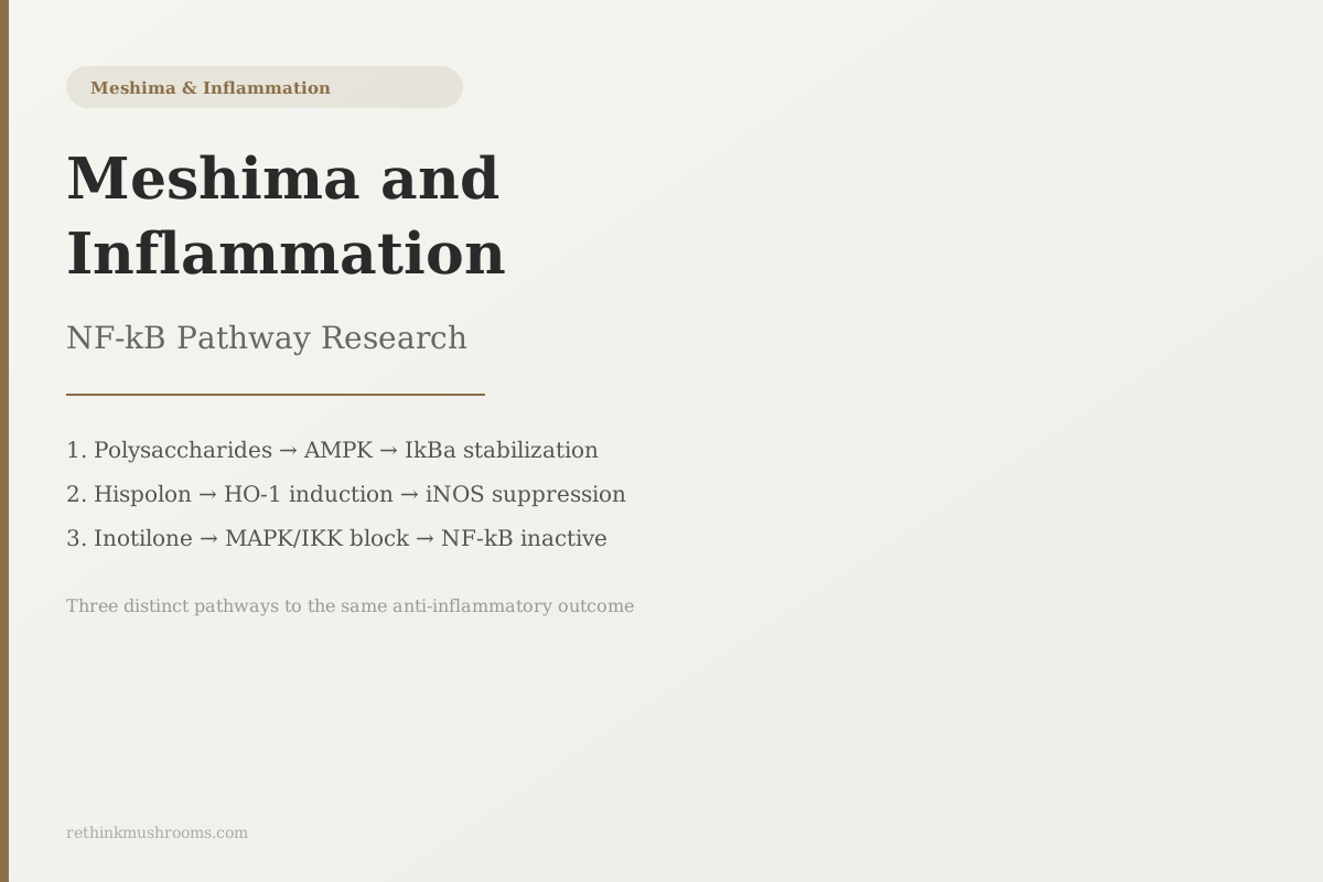
Three distinct Phellinus linteus compounds each suppress NF-kB through separate upstream routes: polysaccharides via AMPK, hispolon via HO-1 induction, and inotilone via MAPK/IKK phosphorylation blocking.
Xie et al., International Journal of Biological Macromolecules, 2019
AMPK
polysaccharides activate this kinase to stabilize IkBa and keep NF-kB inactive (Xie 2019)
HO-1
enzyme induced by hispolon-rich fraction; generates CO and biliverdin that suppress iNOS (Kim 2007)
MAPK/IKK
phosphorylation blocked by inotilone, preventing NF-kB's upstream activation (Huang 2012)
Xie et al., International Journal of Biological Macromolecules, 2019
 View as image ↗
NF-kB is a transcription factor that acts as a master switch for inflammation. When it's activated, it turns on genes for inflammatory cytokines, for iNOS (which produces inflammatory nitric oxide), and for COX-2 (which drives prostaglandin production). Chronically active NF-kB is implicated in arthritis, inflammatory bowel disease, asthma, and cancer progression. Blocking it is a major goal in both pharmaceutical research and the study of natural compounds.
Phellinus linteus (meshima) NF-kB inflammation research spans more than two decades and, unusually for a natural compound, has identified not one but three separate molecular mechanisms by which different parts of the mushroom reach this same pathway from different upstream directions. Each mechanism involves a distinct compound class: polysaccharides, hispolon, and inotilone.
This article maps all three. For the foundational introduction to meshima, see What Is Meshima Mushroom. For the specific compounds responsible for these effects, see Phellinus linteus Bioactive Compounds.
Key Takeaways
- Three distinct Phellinus linteus compounds each suppress NF-kB through separate molecular routes: polysaccharides activate AMPK to stabilize IkBa, hispolon-rich fractions induce HO-1 to suppress iNOS and NF-kB activation, and inotilone blocks MAPK/IKK phosphorylation to inactivate NF-kB upstream
- Xie et al. (2019) found PL polysaccharides activated AMPK in LPS-stimulated macrophages, stabilizing IkBa and simultaneously reducing pro-inflammatory cytokines (TNF-a, IL-1b, IL-6) while increasing anti-inflammatory ones (IL-4, IL-10)
- Huang et al. (2012) found inotilone, a styrenylpyrone compound from PL, blocked MAPK/IKK phosphorylation, suppressed NF-kB activation and MMP-9 expression, and confirmed in vivo anti-inflammatory activity in a carrageenan-induced paw edema model with increased SOD, catalase, and GPx
- Yan et al. (2014) showed oral PL extract suppressed NF-kB and p38 MAPK activity in an asthma mouse model, reducing Th2 cytokines (IL-4, IL-5, IL-13), eosinophil infiltration, and airway inflammation
- A 2023 systematic review by Qin et al. found that NF-kB inhibition potency depends on polysaccharide molecular structure: specific branching patterns and molecular weight determine anti-inflammatory activity, explaining why potency varies across commercial extracts
- All NF-kB inhibition data for Phellinus linteus is preclinical; the only human RCTs (Ku et al. 2022) measured general immune markers, not NF-kB pathway activity or inflammatory cytokines directly
What NF-kB Is and Why Its Inhibition Matters
NF-kB is not a single protein but a family of transcription factors that operate inside nearly every cell type in the body. In its inactive state, it's held in the cytoplasm by a protein called IkBa. When a cell detects a threat (a bacterial signal, oxidative damage, or cytokines from neighboring immune cells), IkBa is phosphorylated and degraded, releasing NF-kB to travel into the nucleus and switch on inflammatory genes.
The genes NF-kB turns on include those for TNF-alpha, IL-1beta, IL-6, iNOS, and COX-2. These are the mediators of the acute inflammatory response: they recruit immune cells, increase vascular permeability, and kill pathogens. In a healthy, transient response, this is necessary. The problem arises when NF-kB becomes chronically active. Sustained NF-kB signaling drives the inflammatory state underlying rheumatoid arthritis, Crohn's disease, asthma, and multiple cancer types.
Volman et al. (2010) demonstrated that mushroom beta-glucan polysaccharides can modulate nitric oxide production and NF-kB transactivation in macrophages, with polysaccharide branching structure determining the magnitude of the effect.[6] This study contextualized mushroom anti-inflammatory research broadly before the compound-specific mechanisms were clarified.
Pathway 1: Polysaccharides via AMPK Activation
The first and most studied NF-kB suppression mechanism in Phellinus linteus involves its polysaccharide fractions activating a cellular energy sensor called AMPK.
AMPK (AMP-activated protein kinase) is the cell's energy checkpoint: it activates when the cell's energy state is low or when it detects specific molecular patterns. When activated, AMPK phosphorylates and stabilizes IkBa, the protein that holds NF-kB in its inactive state. Stable IkBa means NF-kB stays in the cytoplasm, unable to enter the nucleus and switch on inflammatory genes.
Xie et al. (2019) demonstrated this cascade precisely in LPS-stimulated RAW264.7 macrophages, the standard model for studying innate immune inflammation.[1] Phellinus linteus polysaccharides reduced NF-kB nuclear translocation (the step where NF-kB moves from cytoplasm to nucleus) via AMPK activation and IkBa stabilization. The downstream cytokine results were striking: pro-inflammatory cytokines TNF-alpha, IL-1beta, IL-2, IL-6, and IL-12 all decreased, while anti-inflammatory cytokines IL-4 and IL-10 increased. The mushroom's polysaccharides were not simply dampening inflammation uniformly; they were shifting the balance between pro- and anti-inflammatory signaling.
Lin et al. (2014) extended this with fermented Phellinus linteus broths, finding NF-kB suppression accompanied by blockade of MyD88 and TRAF-6, two adapter proteins that transmit inflammatory signals from surface receptors (like TLR4) upstream of NF-kB activation.[3] The finding that the fermentation substrate altered potency compared to standard mycelium is practically relevant: not all PL preparations use the same growing substrate.
For detailed cytokine data including the specific percentage inhibitions from the septic shock model, see Phellinus linteus and Inflammatory Cytokine Regulation.
Pathway 2: Hispolon via HO-1 Induction
The second NF-kB pathway operates through hispolon, a polyphenol compound found in Phellinus linteus, and uses an indirect mechanism that explains an apparent paradox in its biology.
Hispolon is a mild pro-oxidant: it generates reactive oxygen species in cells. Yet it consistently shows anti-inflammatory effects in macrophage models. The resolution lies in a cellular stress response protein called heme oxygenase-1 (HO-1).
Kim et al. (2007) studied a hispolon-rich butanol fraction of Phellinus linteus in LPS-stimulated RAW264.7 macrophages and found it suppressed NF-kB and MAPK activation, iNOS expression, NO production, COX-2 expression, and PGE2 release through redox-based suppression of the signaling cascade.[2] The mechanism: mild oxidative stress from hispolon triggers the cell to induce HO-1 as a protective response. HO-1 then produces carbon monoxide and biliverdin, two molecules with potent anti-inflammatory activity that secondarily suppress JNK phosphorylation and NF-kB activation.
The counterintuitive biology: hispolon uses a small oxidative stress as the signal to activate a more powerful anti-inflammatory defense. The cell interprets the hispolon signal as mild damage and responds by upregulating HO-1, which then suppresses the inflammatory cascade more broadly than hispolon alone could.
This mechanism also extends to neuroinflammation. Research on brain microglial cells has shown hispolon suppresses inflammatory NO production through the same HO-1 route in brain tissue, a finding covered in depth in Meshima Hispolon and iNOS Inhibition.
Pathway 3: Inotilone via MAPK/IKK Phosphorylation Blocking
The third mechanism involves a compound called inotilone, a styrenylpyrone specific to Phellinus linteus, and it operates by blocking the kinase cascade that activates NF-kB before it even reaches IkBa.
IKK (IkB kinase) is the enzyme that phosphorylates IkBa to release NF-kB from its inactive state. If IKK can be blocked, IkBa is never degraded, and NF-kB never moves to the nucleus. MAPK (mitogen-activated protein kinase) is a related upstream kinase that also contributes to the inflammatory cascade. Blocking IKK and MAPK phosphorylation is the most upstream NF-kB inhibition strategy.
Huang et al. (2012) isolated inotilone from Phellinus linteus and tested it in LPS-stimulated macrophages and in a whole-animal carrageenan-induced paw edema model.[4] In vitro, inotilone blocked IKK and MAPK phosphorylation, prevented NF-kB nuclear entry, and suppressed iNOS, MMP-9, and their downstream products (NO, PGE2, and matrix metalloprotease activity). MMP-9 inhibition is separately important: MMPs are enzymes cancer cells use to degrade the extracellular matrix for invasion, making inotilone's mechanism relevant to both inflammation and cancer spread. That angle is explored in Meshima and MMP-9 Inhibition.
In the paw edema mouse model, oral or injected inotilone reduced edema volume and restored antioxidant enzyme levels: superoxide dismutase (SOD), catalase, and glutathione peroxidase (GPx) all increased in treated animals compared to untreated controls. These are the cell's own antioxidant defense proteins. The in vivo data confirms that the in vitro IKK/MAPK inhibition translates to a measurable anti-inflammatory outcome in a living organism.
The Asthma Data: NF-kB Inhibition in Allergic Inflammation
The three cell-culture and septic models above all used LPS, a bacterial cell wall component, to trigger inflammation. Yan et al. (2014) tested a different inflammatory context: allergic airway inflammation in an OVA (ovalbumin) mouse model of asthma, the standard preclinical asthma model.[5]
Oral Phellinus linteus extract significantly inhibited eosinophilic airway inflammation in OVA-challenged mice. Th2 cytokines (IL-4, IL-5, IL-13), eotaxin (an eosinophil-attracting signal), and adhesion molecules were all reduced. The mechanism tracked to downregulation of NF-kB and p38 MAPK activation in the airway tissue.
The asthma data extends the relevance of Phellinus linteus NF-kB research beyond the innate immune activation models. Allergic asthma is a Th2-driven inflammatory condition, distinct from the LPS-triggered innate response in the macrophage studies above. The fact that oral PL extract suppresses NF-kB in both contexts suggests the anti-inflammatory effect is not limited to one type of inflammatory trigger. For more on meshima's effects on chronic inflammatory conditions, see Meshima for Chronic Inflammatory Conditions.
Why Potency Varies Across Studies: Structure-Activity and the Evidence Limits
One of the most important findings from recent reviews concerns why Phellinus linteus anti-inflammatory studies show such variable results across different extracts and preparations.
Qin et al. (2023) conducted a systematic review of PL polysaccharides and found that anti-inflammatory potency is not a fixed property of the mushroom but depends heavily on molecular weight, backbone structure, and branching pattern of the specific polysaccharide fraction.[7] Beta-1,3/1,6-glucan branching patterns produce different activity levels from linear beta-1,3-glucans. High-molecular-weight fractions may perform differently from low-molecular-weight fractions in AMPK activation assays. This explains a consistent frustration in the literature: two studies using "Phellinus linteus" extract may be using structurally different polysaccharides and will not produce comparable results.
Kou et al. (2024) extended this to delivery systems, identifying nanoparticle encapsulation approaches that improve oral bioavailability of PL polysaccharides, which notoriously have low and variable absorption from standard extracts.[8]
The honest limitation: every study in this article used cell culture models or mouse models. NF-kB pathway activity has not been measured in human subjects after Phellinus linteus consumption. The two 2022 RCTs from the same group measured immune markers in healthy adults but did not assess NF-kB, iNOS, cytokine panels, or any of the specific inflammatory endpoints studied in the preclinical research. The mechanistic picture is detailed; the clinical translation gap is substantial. For comparison with another mushroom where NF-kB research exists in a different context, see Enoki and NF-kB Pathway Modulation.
The Bottom Line
Phellinus linteus (meshima) inhibits NF-kB inflammatory signaling through three distinct molecular mechanisms: polysaccharides via AMPK-mediated IkBa stabilization (Xie 2019), hispolon-rich fractions via HO-1 induction that suppresses iNOS and NF-kB (Kim 2007), and inotilone via MAPK/IKK phosphorylation blocking (Huang 2012). In vivo confirmation of anti-inflammatory activity exists in carrageenan paw edema and OVA asthma models. No human trial has directly measured NF-kB or inflammatory cytokine effects after Phellinus linteus supplementation.
Frequently Asked Questions
Sources & References
- 1RTHM-00599·Xie, Zhenglu et al. (2019). “Anti-inflammatory activity of polysaccharides from Phellinus linteus by regulating the NF-κB translocation in LPS-stimulated RAW264.7 macrophages..” International journal of biological macromolecules.Study breakdown →PubMed →↩
- 2RTHM-00116·Kim, Ho Gyoung et al. (2007). “Phellinus linteus inhibits inflammatory mediators by suppressing redox-based NF-kappaB and MAPKs activation in lipopolysaccharide-induced RAW 264.7 macrophage..” Journal of ethnopharmacology.Study breakdown →PubMed →↩
- 3RTHM-00618·Lin, Chun-Jung et al. (2014). “Biological evaluation of Phellinus linteus-fermented broths as anti-inflammatory agents..” Journal of bioscience and bioengineering.Study breakdown →PubMed →↩
- 4RTHM-00460·Huang, Guan-Jhong et al. (2012). “Anti-inflammatory activities of inotilone from Phellinus linteus through the inhibition of MMP-9, NF-κB, and MAPK activation in vitro and in vivo..” PloS one.Study breakdown →PubMed →↩
- 5RTHM-00660·Yan, Guang Hai et al. (2014). “Phellinus linteus Extract Exerts Anti-asthmatic Effects by Suppressing NF-κB and p38 MAPK Activity in an OVA-induced Mouse Model of Asthma..” Immune network.Study breakdown →PubMed →↩
- 6RTHM-00200·Volman, Julia J et al. (2010). “Effects of mushroom-derived beta-glucan-rich polysaccharide extracts on nitric oxide production by bone marrow-derived macrophages and nuclear factor-kappaB transactivation in Caco-2 reporter cells: can effects be explained by structure?.” Molecular nutrition & food research.Study breakdown →PubMed →↩
- 7RTHM-01770·Qin, Dandan et al. (2023). “Polysaccharides from Phellinus linteus: A systematic review of their extractions, purifications, structures and functions..” International journal of biological macromolecules.Study breakdown →PubMed →↩
- 8RTHM-01920·Kou, Fang et al. (2024). “Phellinus linteus polysaccharides: A review on their preparation, structure-activity relationships, and drug delivery systems..” International journal of biological macromolecules.Study breakdown →PubMed →↩