Meshima & Oxidative Stress
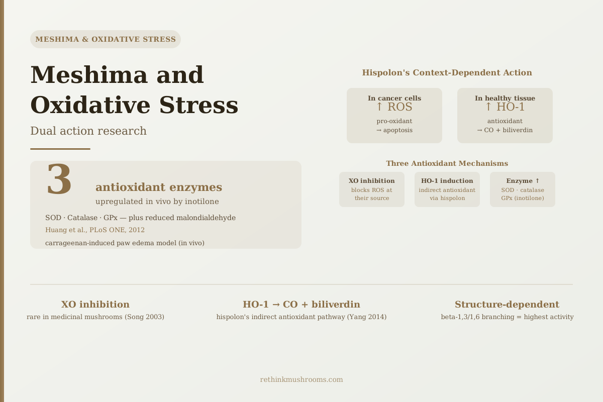
3 enzymes
In a living animal inflammation model, Phellinus linteus inotilone increased all three major antioxidant defense enzymes — SOD, catalase, and GPx — while simultaneously reducing malondialdehyde, the biomarker of oxidative cell damage.
Huang et al., PLoS ONE, 2012
HO-1 induction
hispolon makes macrophages build their own antioxidant enzyme rather than scavenging radicals itself (Yang 2014)
XO inhibition
reported activity against the enzyme that generates reactive oxygen species at source (Song 2003)
Zero human data
no published trial has measured oxidative stress markers in people taking Meshima
Huang et al., PLoS ONE, 2012
 View as image ↗
Antioxidants are commonly described as though they work by catching reactive molecules in the bloodstream — the "free radical scavenger" model. Meshima's relationship with oxidative stress is more complicated and more interesting than that. Research on Phellinus linteus compounds identifies at least four distinct antioxidant mechanisms, and only one of them is scavenging: one blocks the enzyme that produces reactive oxygen species in the first place, one activates the body's own antioxidant enzyme system rather than substituting for it, and one raises the activity of the three core antioxidant enzymes in living tissue.
Adding to the complexity is a finding that is routinely described backwards. Hispolon, Meshima's key polyphenol, is often summarised as "a pro-oxidant in cancer cells and an antioxidant in healthy cells." The cell-culture evidence says something different and more useful: hispolon appears to raise reactive oxygen species in healthy immune cells too — and that oxidative signal is precisely what switches on the protective response. Same action, different outcomes, depending on what the cell can absorb.
This article covers what the oxidative stress research on Meshima has actually found, which mechanisms have the best evidence, and where the evidence stops.
For background on Meshima's general research and its major bioactive compounds, see what Meshima mushroom is and why it's called Korea's anti-cancer mushroom.
Key Takeaways
- Meshima's antioxidant activity operates through at least four distinct mechanisms: direct free radical scavenging, xanthine oxidase inhibition (blocking ROS production at source), HO-1 enzyme induction, and upregulation of the SOD/catalase/GPx enzyme system
- Hispolon induces heme oxygenase-1 (HO-1) in macrophages — an enzyme that generates anti-inflammatory CO and biliverdin — providing protection by upregulating the cell's own defense system rather than by scavenging (Yang 2014)
- The same study found hispolon increased intracellular peroxide in those macrophages, and blocking ROS with N-acetylcysteine prevented HO-1 induction — meaning the oxidative signal is the trigger, not a side effect (Yang 2014)
- Song et al. (2003) reported xanthine oxidase inhibition alongside DPPH scavenging comparable to vitamin C in a Phellinus linteus fruiting-body extract — all measured in vitro
- Huang et al. (2012) tested inotilone in a living inflammation model and found increased SOD, catalase, and GPx alongside reduced malondialdehyde — in vivo antioxidant enzyme upregulation
- Polysaccharide antioxidant potency depends on molecular weight, branching, and extraction method, which is why different Meshima preparations perform differently (Qin 2023, Kou 2024)
- No published human study has measured any oxidative stress marker in people taking Meshima — the entire evidence base is cell culture and animal work
Why Hispolon Raises Oxidative Stress in Healthy Cells Too
Most discussions of antioxidants treat the compound as uniformly protective — it neutralizes reactive molecules wherever it goes. Hispolon doesn't work that way, and the popular description of how it differs is itself inaccurate.
In cancer-cell research, hispolon triggers cell death through pathways involving reactive oxygen species generation — oxidative stress is part of the mechanism by which hispolon kills cancer cells (covered in Meshima and cancer cell apoptosis).
The usual next sentence is that hispolon does the opposite in healthy tissue. It doesn't. Yang and colleagues tested hispolon in macrophages stimulated with LPS, LTA, or peptidoglycan — immune cells under inflammatory attack — and found that hispolon upregulated heme oxygenase-1 (HO-1), an enzyme that protects cells from oxidative damage.[3] But the same experiments recorded an increase in intracellular peroxide in those macrophages. Hispolon was raising oxidative stress in the healthy cells as well.
What makes the finding interpretable is the blocking experiment. When the researchers added N-acetylcysteine — a reactive oxygen species scavenger — HO-1 induction was prevented. Remove the oxidative signal and the protective response never happens. The ROS increase is not an unwanted side effect of hispolon's action in healthy cells; it is the trigger for it.
Fact Check
The most-repeated claim about hispolon is the wrong way round
The claim
“Hispolon acts as a pro-oxidant in cancer cells and as an antioxidant in healthy cells.”
A tidy summary that merges two separate literatures — cancer cell studies and macrophage studies — into a switch that no experiment has demonstrated.
What research shows
Partly TrueIn LPS-stimulated macrophages, hispolon increased intracellular peroxide rather than lowering it. The protective HO-1 response it produced was abolished when a ROS scavenger was added, showing the oxidative signal was required upstream of the protection.
The outcomes really do differ by cell type — protection in macrophages, death in cancer cells. But the difference is in how the cell responds to an oxidative push, not in hispolon switching between pro-oxidant and antioxidant modes. No study has compared the two cell types side by side under matched conditions.
Ref [3] — Yang et al., Journal of Ethnopharmacology, 2014
This reframes the dual action as something better understood than a paradox. A modest oxidative push in a cell with intact defenses provokes an adaptive response that leaves the cell better protected than before — the pattern toxicology calls hormesis. The same push in a cell already running near its antioxidant limit, as many cancer cells do, exhausts those defenses instead. The compound behaves consistently; the cells do not.
Two limits are worth stating plainly. All of this is cell culture, at concentrations researchers chose to elicit a response. And no experiment has tested cancer cells and healthy cells side by side under matched conditions, so the selectivity is inferred from separate literatures rather than demonstrated in one comparison.
Mechanism chain
Hispolon Protects Macrophages By Stressing Them First
The popular summary has hispolon lowering oxidative stress in healthy cells. In the study most often cited for that claim, it raised it — and the increase was necessary for the protective enzyme to appear. Below is the chain as reported, followed by the blocking experiments that establish the order of events.
Hispolon enters a stimulated macrophage
Oxidative pushRAW264.7 cells challenged with LPS, LTA, or peptidoglycan — immune cells already under inflammatory load
Intracellular peroxide rises
Oxidative pushMeasured by DCHF-DA staining. Oxidative stress goes up, not down — in the healthy cell
HO-1 is transcribed and translated
Adaptive responseHeme oxygenase-1 increases at both mRNA and protein level — the cell builds new defensive enzyme
iNOS and nitric oxide fall
Adaptive responseConcentration-dependent suppression of iNOS protein and NO output, alongside reduced JNK, AP-1, and NF-kB activity
The macrophage survives the inflammatory hit
OutcomeHispolon protected the cells from LPS-, LTA-, and PGN-induced apoptosis
How the order of events was established
Each agent removes one step. If HO-1 still rises, that step was not required. All three blocked it.
N-acetylcysteine
Scavenges reactive oxygen species→ HO-1 induction prevented
The ROS rise is required upstream — remove the oxidative signal and no protection develops
Actinomycin D
Gene transcription→ HO-1 increase prevented
New HO-1 mRNA is being made — this is gene activation
Cycloheximide
Protein translation→ HO-1 increase prevented
New enzyme is being synthesised, not released from a stored pool
Why the same compound can kill a cancer cell
Nothing above shows hispolon switching modes. It pushes cells toward oxidative stress; what differs is whether the cell can answer. A macrophage with intact reserves answers by building HO-1 and ends up better defended than it started — the pattern toxicology calls hormesis. Cancer cells, which frequently operate closer to their antioxidant ceiling, are reported in a separate cell-culture literature to undergo apoptosis under the same kind of push. That is an inference across two sets of experiments, not a measured comparison: no study has run both cell types side by side under matched conditions, and all of this work is in culture, at concentrations chosen to produce a response.
Chain and blocking experiments taken from the published abstract of ref [3]
Xanthine Oxidase Inhibition: An Uncommon Mechanism in Mushroom Research
One of the more mechanistically specific antioxidant findings in the Meshima literature is xanthine oxidase inhibition.
Song and colleagues published an early characterization of Phellinus linteus in the Journal of Ethnopharmacology in 2003, testing a 70% ethanol extract of the fruiting bodies.[1] The extract scavenged the stable free radical DPPH at a level the authors described as comparable to vitamin C, and inhibited lipid peroxidation in a concentration-dependent way. Alongside those scavenging results, and alongside anti-angiogenic activity in a chick embryo assay, the study reported inhibition of xanthine oxidase — an enzyme that catalyzes the conversion of hypoxanthine to xanthine to uric acid, generating superoxide and hydrogen peroxide as byproducts.
Xanthine oxidase matters in two contexts. During inflammation, activated immune cells produce large quantities of xanthine oxidase-derived ROS as part of the oxidative burst that damages tissue alongside pathogens. And in gout and other conditions of purine overload, excessive xanthine oxidase activity produces both excessive uric acid and excessive oxidative stress at once.
Blocking xanthine oxidase addresses oxidative stress at its source rather than downstream. Instead of neutralizing ROS after generation, XO inhibition reduces how many are produced. The best-known clinical XO inhibitor is allopurinol, used for gout. It is worth being careful with that comparison: Song's study measured enzyme inhibition in vitro and did not compare potency against allopurinol or test whether the effect occurs at doses achievable from a supplement. Shared enzyme activity is a mechanistic starting point, not evidence of comparable clinical effect.
XO inhibition is also not among the mechanisms most prominently reported for reishi, lion's mane, turkey tail, or shiitake, which makes it a distinguishing feature of the Meshima literature. That reflects what researchers have chosen to measure and report, though, rather than a head-to-head comparison establishing that other species lack the activity.
HO-1 Induction: How Hispolon Activates the Cell's Internal Defense
The Yang 2014 study is the clearest illustration of how hispolon's antioxidant mechanism works in healthy immune cells, and it is worth understanding in detail because it is counterintuitive.
Heme oxygenase-1 (HO-1) is an enzyme that breaks down heme — the iron-containing component of hemoglobin — into three products: carbon monoxide (CO), biliverdin, and free iron. This sounds like a destructive process, but CO and biliverdin are both anti-inflammatory: CO blocks inflammatory signaling in smooth muscle and immune cells, and biliverdin (which converts to bilirubin) is a potent antioxidant that scavenges reactive oxygen species.
Yang and colleagues showed that hispolon upregulates HO-1 in stimulated macrophages at both the protein and mRNA level, via a pathway involving suppression of JNK phosphorylation and of NF-kB and AP-1 activation — key drivers of inflammatory gene expression.[3] The net effect: reduced iNOS expression and nitric oxide production, protection against inflammation-induced apoptosis, and an overall shift toward anti-inflammatory cell behavior.
That the induction was blocked by cycloheximide and actinomycin D — inhibitors of protein translation and gene transcription respectively — confirms the cell was genuinely manufacturing new HO-1 rather than releasing a stored supply.
Indirect mechanisms are potentially more durable than direct scavenging. Once HO-1 is upregulated, the cell continues producing CO and biliverdin for as long as the enzyme remains active, rather than requiring continuous delivery of a scavenging compound. Whether that durability survives digestion, absorption, and distribution at supplement doses is a separate question these experiments do not address.
For a deeper look at how hispolon specifically inhibits iNOS through the HO-1 pathway and what this means for inflammatory conditions, see Meshima hispolon and iNOS inhibition: a targeted anti-inflammatory mechanism.
In Vivo Antioxidant Evidence: What a Carrageenan Inflammation Model Showed
Cell culture studies establish whether a mechanism is possible. Animal models test whether it actually occurs in living biology. For Meshima's antioxidant mechanisms, the most relevant in vivo data comes from a 2012 study by Huang and colleagues published in PLoS ONE.
Huang et al. (2012) · PLoS ONE
- Population
- Mice with carrageenan-induced hind paw edema, with parallel work in LPS-stimulated RAW264.7 mouse macrophages
- Intervention
- Inotilone, a phenolic compound isolated from Phellinus linteus — chemically distinct from hispolon
- Finding
- Inotilone reduced paw edema and attenuated malondialdehyde in the inflamed paw, while increasing catalase, superoxide dismutase, and glutathione peroxidase activity. It also lowered serum nitric oxide and TNF-alpha, and blocked iNOS, NF-kB, and MMP-9 expression in macrophages.
positive A well-constructed animal study, but a single acute inflammation model in mice. Inotilone is not the compound most Meshima supplements are standardized to, and no dose translation to humans has been established.
Ref [4] — Huang et al., PLoS ONE, 2012
Huang's team isolated inotilone from Phellinus linteus and tested it in a carrageenan-induced paw edema model in mice.[4] Carrageenan injection triggers a defined inflammatory response with both inflammatory signaling and oxidative stress components, making it a standard model for testing anti-inflammatory and antioxidant compounds in vivo.
The antioxidant findings: inotilone attenuated malondialdehyde levels in inflamed tissue — malondialdehyde is produced when lipid membranes are oxidatively damaged, so its reduction signals less oxidative cell damage occurring. Simultaneously, inotilone increased superoxide dismutase (SOD), catalase, and glutathione peroxidase (GPx) activity. These are the three core enzymes of the primary antioxidant defense system: SOD converts superoxide radicals to hydrogen peroxide, and catalase and GPx break that hydrogen peroxide down to water.
The simultaneous increase in all three antioxidant enzymes alongside the decrease in the oxidative damage marker is an internally consistent set of results. It suggests inotilone was activating enzymatic antioxidant defenses — the same broad strategy hispolon uses via HO-1 — rather than only neutralizing free radicals directly.
This is the strongest antioxidant evidence Meshima has, and it is worth being clear about what it is: one acute inflammation model, in mice, using an isolated compound that consumer Meshima products are not standardized to contain.
How Beta-Glucan Structure Determines Antioxidant Potency
The polysaccharide fraction of Meshima also carries antioxidant activity, and this is where structural biology becomes practically relevant.
Volman and colleagues tested beta-glucan-rich polysaccharide extracts from several mushrooms, including Phellinus linteus, in 2010, measuring nitric oxide production by mouse bone marrow-derived macrophages and NF-kB transactivation in human intestinal Caco-2 reporter cells.[2] Their structural analysis indicated that branching of the beta-glucan chain is essential for immune-stimulating activity. Notably, the Phellinus linteus extract they tested produced no effect at all on nitric oxide production, while extracts from Agaricus bisporus did — a direct demonstration that "mushroom beta-glucan" is not a single functional ingredient, and that a Phellinus preparation can be inert in an assay where another species is active. That study measured immune signaling endpoints rather than antioxidant ones, so it speaks to structure-dependence generally rather than to Meshima's antioxidant potency specifically.
Xie and colleagues in 2019 showed that Phellinus linteus polysaccharides reduced NF-kB nuclear translocation, associated with decreased IkBa phosphorylation and increased AMPK phosphorylation, while shifting the cytokine balance: decreasing TNF-alpha, IL-1beta, IL-2, IL-6, and IL-12 while increasing anti-inflammatory IL-4 and IL-10.[5] That study measured cytokines and NF-kB rather than oxidative stress markers directly. The antioxidant relevance is inferred: inflammatory cytokines upregulate ROS-generating enzymes in immune cells, so suppressing them should reduce oxidative load. That inference is reasonable but was not tested in this experiment.
The 2023 systematic review by Qin and colleagues synthesized structure-activity data across Phellinus linteus polysaccharide studies, reporting that their structure-activity relationships are closely tied to anti-oxidation and anti-tumor activity, and that extraction and purification conditions shape the result.[6] The 2024 review by Kou and colleagues reached a similar conclusion — that bioactivity varies with the complex structure of these polysaccharides, which currently limits their application — and surveyed nano-carrier delivery approaches aimed at improving it.[7]
The practical implication is that "Meshima antioxidant activity" is not a single fixed value. It varies with how the mushroom was processed, which polysaccharide fractions were preserved, and at what molecular weight. Products reporting only milligrams of Phellinus linteus per serving, without polysaccharide standardization, omit the information needed to assess antioxidant potency — and the Volman result is a reminder that a preparation can test inert.
For how Meshima's polysaccharide and hispolon compounds relate to each other structurally and functionally, see Phellinus linteus bioactive compounds: polysaccharides and phenols. For a comparison of antioxidant approaches across medicinal mushrooms, see ergothioneine and chronic inflammatory conditions.
Where the Evidence Stands and What's Missing
The antioxidant research for Meshima is internally consistent and mechanistically specific: direct radical scavenging and xanthine oxidase inhibition from the crude extract, HO-1 induction by hispolon, inotilone upregulation of SOD, catalase, and GPx in a living animal, and polysaccharide-driven NF-kB suppression that plausibly lowers cytokine-driven oxidative load. Four distinct compounds or fractions, four mechanistic routes.
- Cell studiescompleted
Extract scavenging and xanthine oxidase inhibition (Song 2003); hispolon HO-1 induction in macrophages (Yang 2014); polysaccharide NF-kB and cytokine effects (Xie 2019).
- Animal studiescurrent
One study: inotilone raised SOD, catalase, and GPx and lowered malondialdehyde in a mouse carrageenan paw edema model (Huang 2012).
- Small human trialsmissing
Two 2022 randomised trials of Phellinus linteus extract exist, but they measured immune markers — NK cell activity, cytokines, immunoglobulins — and no oxidative stress endpoint.
- Large RCTsmissing
None conducted.
- Meta-analysismissing
Not possible without human oxidative stress trials to pool.
Meshima's antioxidant mechanisms are well characterised in cells and demonstrated once in mice. The human rung is empty: no published trial has measured malondialdehyde, 8-OHdG, F2-isoprostanes, or any other oxidative stress marker in people taking Meshima.
Refs [1]–[7]; human trial scope from refs [8] and [9]
What is absent is any human study measuring Meshima's effect on oxidative stress markers. Markers like malondialdehyde, 8-hydroxy-2'-deoxyguanosine (8-OHdG), or F2-isoprostanes are standard clinical indicators of oxidative cell damage, and none has been tested.
The closest human evidence comes from two 2022 randomised, double-blind, placebo-controlled trials of Phellinus linteus extract by Ku and colleagues. In the larger trial, 98 participants took 1,000 mg/day of extract or dextrin placebo for eight weeks; natural killer cell activity, the primary endpoint, increased significantly versus placebo, while the secondary cytokine and immunoglobulin endpoints did not reach significance.[8] The second was a 30-person pilot in which NK cell activity and IL-6 tended to improve without reaching statistical significance.[9] These are not two independent replications — the same authors led both, in the same journal, months apart — and neither measured an oxidative stress endpoint. The honest summary is one significant immune biomarker in one trial, not confirmed antioxidant activity in humans.
Safety
ModerateAntioxidant supplements and cancer treatment are a genuine interaction question
Concern
Much of Meshima's research sits in cancer biology, and hispolon's cancer-cell activity depends on driving oxidative stress upward. Radiotherapy and several chemotherapy drugs also work partly by generating reactive oxygen species. Whether a compound that raises antioxidant enzyme capacity in healthy tissue could blunt those treatments — or add to them — has not been tested in people.
What the research says
No clinical study has evaluated Meshima alongside cancer treatment. Both the interference scenario and the enhancement scenario are extrapolations from cell and animal work, and neither should be treated as established.
Particularly relevant for: Anyone undergoing or preparing for chemotherapy or radiotherapy, and anyone taking allopurinol or other urate-lowering therapy given the xanthine oxidase overlap
What to do
Discuss any medicinal mushroom supplement with your oncology or prescribing team before starting. This article summarises preclinical research and is not medical advice.
Interaction rationale from refs [1] and [3]; no clinical data exists on this question
The gap between "improved one immune marker" and "reduced oxidative damage" is the next research question, and it is a straightforward one to answer — the oxidative stress markers are inexpensive, standardised, and could be added as secondary endpoints to a trial of the kind already run twice.
For the broader anti-inflammatory picture across chronic conditions including IBD and asthma, see Meshima for chronic inflammatory conditions. For the NF-kB pathway research that connects the antioxidant and anti-inflammatory mechanisms, see Meshima and inflammation: NF-kB pathway research.
The Bottom Line
Meshima's antioxidant activity operates through multiple mechanisms beyond direct free radical scavenging: xanthine oxidase inhibition blocks ROS production at the source; hispolon induces HO-1 so the cell generates its own anti-inflammatory CO and biliverdin; inotilone upregulates SOD, catalase, and GPx in a mouse model. Polysaccharide potency depends on molecular weight, branching, and extraction method, so preparations differ — one tested Phellinus extract produced no effect at all in an immune assay. The widely repeated claim that hispolon is a pro-oxidant in cancer cells and an antioxidant in healthy ones is the wrong way round: it raised oxidative stress in healthy macrophages too, and that signal was required for the protective response. The entire evidence base is cell culture and animal work — no human study has measured an oxidative stress marker in people taking Meshima.
Frequently Asked Questions
Sources & References
- 1RTHM-00121·Song, Yun Seon et al. (2003). “Anti-angiogenic, antioxidant and xanthine oxidase inhibition activities of the mushroom Phellinus linteus..” Journal of ethnopharmacology.Study breakdown →PubMed →↩
- 2RTHM-00200·Volman, Julia J et al. (2010). “Effects of mushroom-derived beta-glucan-rich polysaccharide extracts on nitric oxide production by bone marrow-derived macrophages and nuclear factor-kappaB transactivation in Caco-2 reporter cells: can effects be explained by structure?.” Molecular nutrition & food research.Study breakdown →PubMed →↩
- 3RTHM-00662·Yang, Liang-Yo et al. (2014). “Hispolon inhibition of inflammatory apoptosis through reduction of iNOS/NO production via HO-1 induction in macrophages..” Journal of ethnopharmacology.Study breakdown →PubMed →↩
- 4RTHM-00460·Huang, Guan-Jhong et al. (2012). “Anti-inflammatory activities of inotilone from Phellinus linteus through the inhibition of MMP-9, NF-κB, and MAPK activation in vitro and in vivo..” PloS one.Study breakdown →PubMed →↩
- 5RTHM-00599·Xie, Zhenglu et al. (2019). “Anti-inflammatory activity of polysaccharides from Phellinus linteus by regulating the NF-κB translocation in LPS-stimulated RAW264.7 macrophages..” International journal of biological macromolecules.Study breakdown →PubMed →↩
- 6RTHM-01770·Qin, Dandan et al. (2023). “Polysaccharides from Phellinus linteus: A systematic review of their extractions, purifications, structures and functions..” International journal of biological macromolecules.Study breakdown →PubMed →↩
- 7RTHM-01920·Kou, Fang et al. (2024). “Phellinus linteus polysaccharides: A review on their preparation, structure-activity relationships, and drug delivery systems..” International journal of biological macromolecules.Study breakdown →PubMed →↩
- 8RTHM-01556·Ku, Yong Ho et al. (2022). “Effects of Phellinus linteus extract on immunity improvement: A CONSORT-randomized, double-blinded, placebo-controlled trial..” Medicine.Study breakdown →PubMed →↩
- 9RTHM-01555·Ku, Yong Ho et al. (2022). “Efficacy of Phellinus linteus extract on immunity enhancement: A CONSORT-randomized, double-blind, placebo-controlled pilot trial..” Medicine.Study breakdown →PubMed →↩