Agaricus blazei and Diabetes
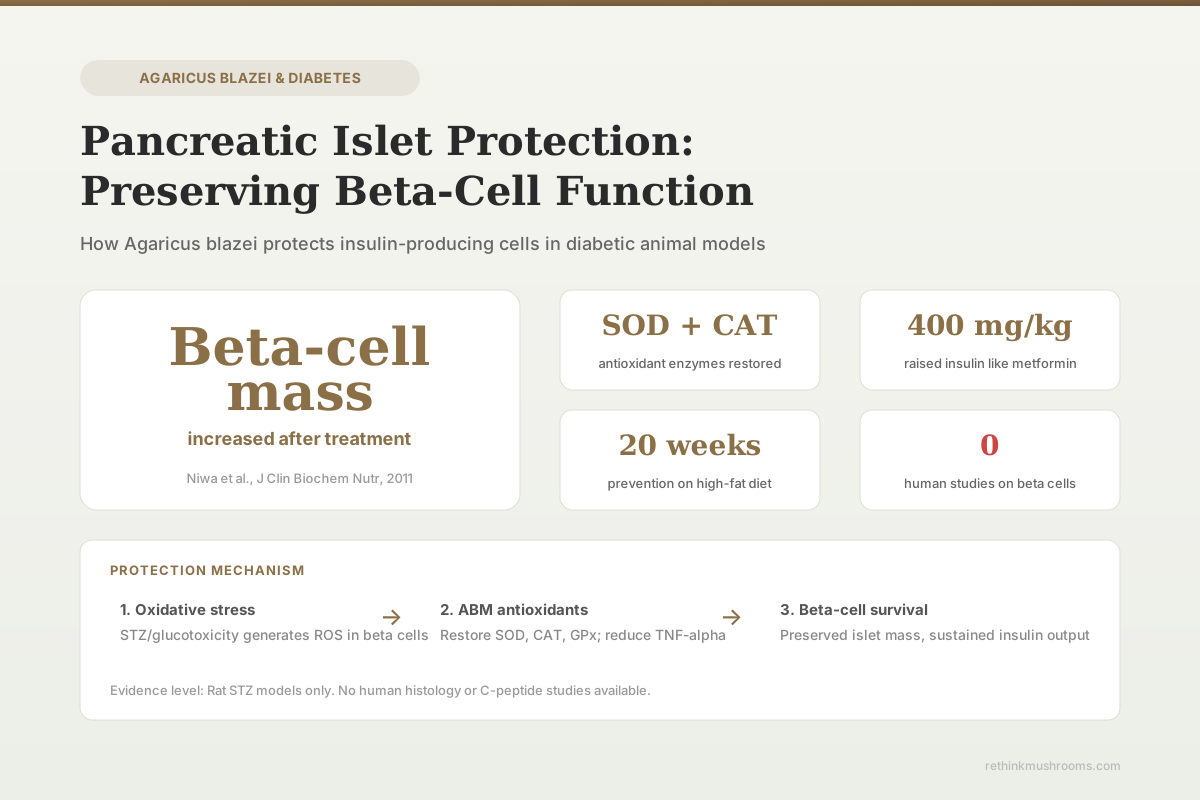
Beta-cell mass increased
Histological examination showed Agaricus blazei treatment increased pancreatic beta-cell mass in streptozotocin-induced diabetic rats after two months of daily oral feeding.
Niwa et al., Journal of Clinical Biochemistry and Nutrition, 2011
SOD, CAT, GPx
restored in pancreatic tissue
Plasma insulin
elevated dose-dependently by EAF
0 human studies
on beta-cell protection specifically
Niwa et al., Journal of Clinical Biochemistry and Nutrition, 2011
 View as image ↗
Agaricus blazei beta-cell protection research comes from a handful of animal studies that measured what happens to insulin-producing cells when diabetic rats receive ABM extracts. The findings are consistent: beta-cell mass increases, antioxidant enzymes recover, and oxidative damage markers decline.[7] For the broader picture of how these pancreatic effects relate to blood sugar lowering, see our overview of the hypoglycemic evidence.
No human study has directly measured ABM's effects on pancreatic beta cells. The human evidence for ABM and diabetes measures insulin resistance (HOMA-IR) and adiponectin, not islet cell histology. Everything in this article comes from rat models, primarily using streptozotocin (STZ) to destroy beta cells chemically.
Key Takeaways
- ABM treatment increased pancreatic beta-cell mass in STZ-induced diabetic rats as shown by aldehyde fuchsin staining of islets (Niwa et al., 2011)
- ABM ethyl acetate extract restored superoxide dismutase, catalase, and glutathione peroxidase in pancreatic tissue of diabetic rats (Wei et al., 2020)
- The ethyl acetate fraction elevated plasma insulin levels dose-dependently in STZ rats, with 400 mg/kg matching metformin at 500 mg/kg (Oh et al., 2010)
- ABM dietary supplementation prevented insulin resistance in rats fed a high-fat diet for 20 weeks, before beta-cell damage occurred (Vincent et al., 2013)
- Oxidative stress suppression and reduced TNF-alpha production preceded beta-cell mass improvement, suggesting antioxidant protection as the primary mechanism (Niwa et al., 2011)
- No human study has examined ABM's direct effects on pancreatic beta-cell function or mass
Why Beta Cells Matter in Diabetes
Pancreatic beta cells, clustered in the islets of Langerhans, are the body's sole source of insulin. In type 1 diabetes, autoimmune destruction eliminates most beta cells. In type 2 diabetes, chronic insulin resistance places sustained demand on beta cells, which eventually exhaust and lose function. By the time type 2 diabetes is diagnosed, an estimated 50% of beta-cell function has already been lost.[8]
Preserving remaining beta cells is a central goal of diabetes management. Once beta cells die, they do not regenerate in adults (despite some animal evidence of limited proliferation). Any intervention that slows beta-cell destruction could extend the period during which the body maintains some endogenous insulin production, potentially delaying the progression from oral medications to injectable insulin.
This is the therapeutic context for ABM's beta-cell research. If ABM genuinely protects beta cells from oxidative destruction, it could have value beyond simply lowering blood glucose. For the clinical evidence in type 2 diabetes, that question remains unanswered.
Direct Evidence: Beta-Cell Mass Measurement
Niwa et al. (2011): Histological Confirmation
Niwa et al. (2011) provided the most direct evidence for beta-cell protection.[2] STZ-induced diabetic rats received daily oral ABM powder for two months. Pancreatic tissue was then examined histologically using aldehyde fuchsin staining, which specifically stains insulin-containing beta-cell granules purple or blue.
The results showed a clear gradient. Untreated diabetic rats had severely degranulated islets with few remaining beta cells, the expected outcome after STZ destruction. ABM-treated diabetic rats showed measurably increased beta-cell mass compared to untreated diabetic controls. The islets appeared larger and retained more stained granules, indicating more functional insulin-producing cells.
This study used ABM in combination with Ipomoea batatas (sweet potato) extract, which complicates attribution. Both compounds individually and in combination were tested, and ABM alone did show beta-cell improvement, but the combination produced the strongest effects. The study also found reduced superoxide production and lower vascular nitrotyrosine formation, suggesting that oxidative stress suppression preceded the beta-cell recovery.
The limitation: STZ destroys beta cells through a specific mechanism (DNA alkylation via GLUT2 uptake). The "recovery" seen could represent protection of surviving beta cells from ongoing oxidative damage rather than true regeneration of destroyed cells. The study cannot distinguish between these possibilities.
Wei et al. (2020): Pancreatic Tissue Recovery
Wei et al. (2020) focused on the antioxidant enzyme recovery in pancreatic tissue.[1] STZ-induced diabetic rats received either ABM ethanol extract or ethyl acetate extract. Both were compared against untreated diabetic controls.
The ethyl acetate extract was more effective. It restored superoxide dismutase (SOD), catalase (CAT), and glutathione peroxidase (GPx) levels in pancreatic tissue toward normal values. AST and ALT also improved. Histopathological analysis confirmed pancreatic tissue structural improvement.
This study links the beta-cell protection to a specific mechanism: antioxidant enzyme restoration. STZ generates reactive oxygen species in beta cells as part of its destructive mechanism. By rebuilding the antioxidant defense system in pancreatic tissue, ABM extracts could shield surviving beta cells from the secondary oxidative cascade that continues after the initial STZ insult. This same liver protection mechanism appears to operate in pancreatic tissue.
Indirect Evidence: Insulin Level Recovery
Oh et al. (2010): Plasma Insulin and GLUT-4
Oh et al. (2010) measured plasma insulin levels as an indirect marker of beta-cell function.[3] The ethyl acetate fraction of ABM submerged-culture broth elevated plasma insulin in STZ-diabetic rats in a dose-dependent manner over 14 days. At 400 mg/kg, the insulin elevation was comparable to the glucose-lowering achieved by metformin at 500 mg/kg.
Higher plasma insulin in STZ-treated animals implies either preserved beta-cell mass (more cells producing insulin) or increased per-cell insulin secretion (remaining cells working harder). The study did not perform histology, so it cannot distinguish between these. However, the dose-dependent relationship suggests a graduated biological effect rather than an all-or-nothing threshold.
The study also found increased GLUT-4 protein expression in muscle tissue, which is a peripheral insulin sensitivity effect rather than a beta-cell effect. The combination of increased insulin production and improved insulin sensitivity could explain the metformin-comparable glucose lowering through a fundamentally different mechanism: metformin works primarily on hepatic glucose production and peripheral sensitivity, while ABM may additionally support the insulin supply side.
Di Naso et al. (2010): Systemic Oxidative Stress
Di Naso et al. (2010) measured oxidative stress in the pulmonary tissue of STZ-diabetic rats treated with ABM.[4] While this study examined lungs, not pancreas, it demonstrates that ABM's antioxidant effects are systemic. If ABM reduces oxidative stress throughout the body, the pancreas would benefit alongside other organs.
This systemic antioxidant effect is relevant because diabetic beta-cell damage is not only caused by the initial insult (whether autoimmune attack in type 1 or glucotoxicity in type 2). Chronic systemic inflammation and oxidative stress in diabetes create an environment that continuously damages surviving beta cells. An intervention that reduces this background oxidative load could slow the progressive loss of beta-cell function that characterizes type 2 diabetes progression.
Prevention vs. Treatment: The Vincent Model
Vincent et al. (2013) tested a fundamentally different scenario: can ABM prevent beta-cell stress from developing in the first place?[5] Rats fed a high-fat diet for 20 weeks developed obesity and insulin resistance. Those receiving concurrent ABM supplementation maintained better insulin sensitivity and accumulated less visceral fat.
This study did not measure beta-cell mass directly. But insulin sensitivity preservation means beta cells face less compensatory demand. In the high-fat diet model, beta cells fail because they cannot keep up with the insulin resistance driven by obesity and ectopic fat deposition. By preventing insulin resistance, ABM may protect beta cells from the exhaustion that leads to type 2 diabetes.
This prevention framework is arguably more relevant to human type 2 diabetes than the STZ treatment models. Most humans at risk for type 2 diabetes have functioning beta cells under increasing metabolic stress, not chemically destroyed beta cells. The high-fat diet model recapitulates this trajectory: progressive weight gain drives insulin resistance, which forces beta cells to produce more insulin, which eventually exhausts them. By intervening at the insulin resistance stage, ABM may break this cycle before irreversible beta-cell damage occurs.
The clinical parallel would be prediabetes, a condition affecting an estimated 98 million American adults. In prediabetes, beta cells are working overtime to compensate for rising insulin resistance but have not yet failed. If ABM's insulin-sensitizing effects translate to humans, the greatest benefit might be in this pre-failure window rather than in established diabetes where significant beta-cell loss has already occurred. For how this relates to insulin sensitivity improvements, see our dedicated analysis.
The Mechanism: Antioxidant Shield
Across all studies, the mechanism converges on oxidative stress reduction. Beta cells are particularly vulnerable to oxidative damage because they express relatively low levels of antioxidant enzymes compared to other cell types. This makes them disproportionately sensitive to reactive oxygen species.
In the STZ model, STZ enters beta cells through GLUT2 transporters and generates free radicals that damage DNA, lipid membranes, and proteins. The initial destruction is rapid, but a secondary wave of oxidative damage continues in surviving cells. ABM's antioxidant compounds, including polysaccharides, phenolic compounds, and ergosterol, appear to neutralize this secondary wave.
The evidence suggests ABM's protective effect on beta cells is primarily about defense, not regeneration. The mushroom's compounds restore antioxidant enzyme activity in pancreatic tissue, reduce lipid peroxidation, and lower pro-inflammatory cytokines (particularly TNF-alpha). These effects preserve surviving beta cells rather than creating new ones.
Shamim et al. (2023) reviewed the broader evidence for edible mushroom polysaccharides in diabetes and noted that multiple mushroom species show similar beta-cell protective effects, suggesting this is a property of fungal polysaccharides generally rather than unique to ABM.[6] Ganoderma lucidum (reishi), Grifola frondosa (maitake), and Agaricus bisporus (common button mushroom) all demonstrate antidiabetic effects in various models. Whether ABM is more or less effective than other medicinal mushrooms for beta-cell protection has not been compared directly. This broader pattern suggests the beta-glucan polysaccharides shared across these species may be the primary active compounds, with species-specific secondary metabolites providing additional effects.
What This Evidence Cannot Tell Us
Every study in this article used the STZ rat model, which has a specific limitation for beta-cell research: STZ destroys beta cells through a mechanism unrelated to human type 2 diabetes. In humans, beta cells fail from chronic glucolipotoxicity, endoplasmic reticulum stress, and inflammatory signaling, not from acute chemical destruction. The oxidative stress component overlaps, but the primary drivers differ.
No study has measured beta-cell function in humans taking ABM. The Hsu et al. (2007) diabetes trial measured HOMA-IR and adiponectin but not C-peptide, proinsulin, or beta-cell mass.[7] C-peptide, a byproduct of insulin production, is the standard human biomarker for beta-cell function. Until a study measures C-peptide response in humans taking ABM, the beta-cell protection claim remains confined to rat histology.
The distinction between protection and regeneration remains unresolved. The histological evidence shows more beta-cell mass after ABM treatment, but "more" compared to untreated STZ rats could mean either preserved surviving cells or actual proliferation. Distinguishing these would require proliferation markers (Ki-67 staining) that none of the current studies performed.
The dose-response relationship also lacks clarity. Oh et al. (2010) showed dose-dependent insulin elevation with the ethyl acetate fraction, but no study has established a dose-response curve for beta-cell mass preservation. Whether higher doses produce more protection, and at what point diminishing returns or toxicity emerge, remain open questions. The drug interaction profile of ABM adds complexity: any beta-cell protective intervention would need to be compatible with existing diabetes medications, and the CYP enzyme interactions and additive hypoglycemic effects create unanswered safety questions for combination use. The dosage considerations for potential beta-cell protection remain entirely speculative without human dose-finding studies.
The Bottom Line
Agaricus blazei increases beta-cell mass and restores pancreatic antioxidant enzymes in STZ-induced diabetic rats. The mechanism centers on oxidative stress reduction: restoring SOD, catalase, and GPx while suppressing TNF-alpha. A prevention model also shows ABM preserves insulin sensitivity before beta-cell exhaustion occurs. All evidence comes from rat studies using chemical diabetes models, and no human study has measured ABM's effects on beta-cell function directly.
Frequently Asked Questions
Sources & References
- 1RTHM-00661·Wei, Qi et al. (2020). “The beneficial effects of Agaricus blazei Murrill on hepatic antioxidant enzymes and the pancreatic tissue recovery in streptozotocin-induced diabetic rats..” Journal of food biochemistry.Study breakdown →PubMed →↩
- 2RTHM-00227·Niwa, Atsuko et al. (2011). “Ipomoea batatas and Agarics blazei ameliorate diabetic disorders with therapeutic antioxidant potential in streptozotocin-induced diabetic rats..” Journal of clinical biochemistry and nutrition.Study breakdown →PubMed →↩
- 3RTHM-00195·Oh, Tae W et al. (2010). “Semipurified fractions from the submerged-culture broth of Agaricus blazei Murill reduce blood glucose levels in streptozotocin-induced diabetic rats..” Journal of agricultural and food chemistry.Study breakdown →PubMed →↩
- 4RTHM-00179·Di Naso, Fábio Cangeri et al. (2010). “Effect of Agaricus blazei Murill on the pulmonary tissue of animals with streptozotocin-induced diabetes..” Experimental diabetes research.Study breakdown →PubMed →↩
- 5RTHM-00308·Vincent, Mylène et al. (2013). “Dietary supplementation with Agaricus blazei murill extract prevents diet-induced obesity and insulin resistance in rats..” Obesity (Silver Spring.Study breakdown →PubMed →↩
- 6RTHM-00850·Shamim, Mohammad Zaki et al. (2023). “Exploring Edible Mushrooms for Diabetes: Unveiling Their Role in Prevention and Treatment..” Molecules (Basel.Study breakdown →PubMed →↩
- 7RTHM-00114·Hsu, Chung-Hua et al. (2007). “The mushroom Agaricus Blazei Murill in combination with metformin and gliclazide improves insulin resistance in type 2 diabetes: a randomized, double-blinded, and placebo-controlled clinical trial..” Journal of alternative and complementary medicine (New York.Study breakdown →PubMed →↩
- 8RTHM-00765·Huang, Kaiyuan et al. (2022). “Critical review on chemical compositions and health-promoting effects of mushroom Agaricus blazei Murill..” Current research in food science.Study breakdown →PubMed →↩