Ergothioneine & Parkinson's Disease
3 model systems
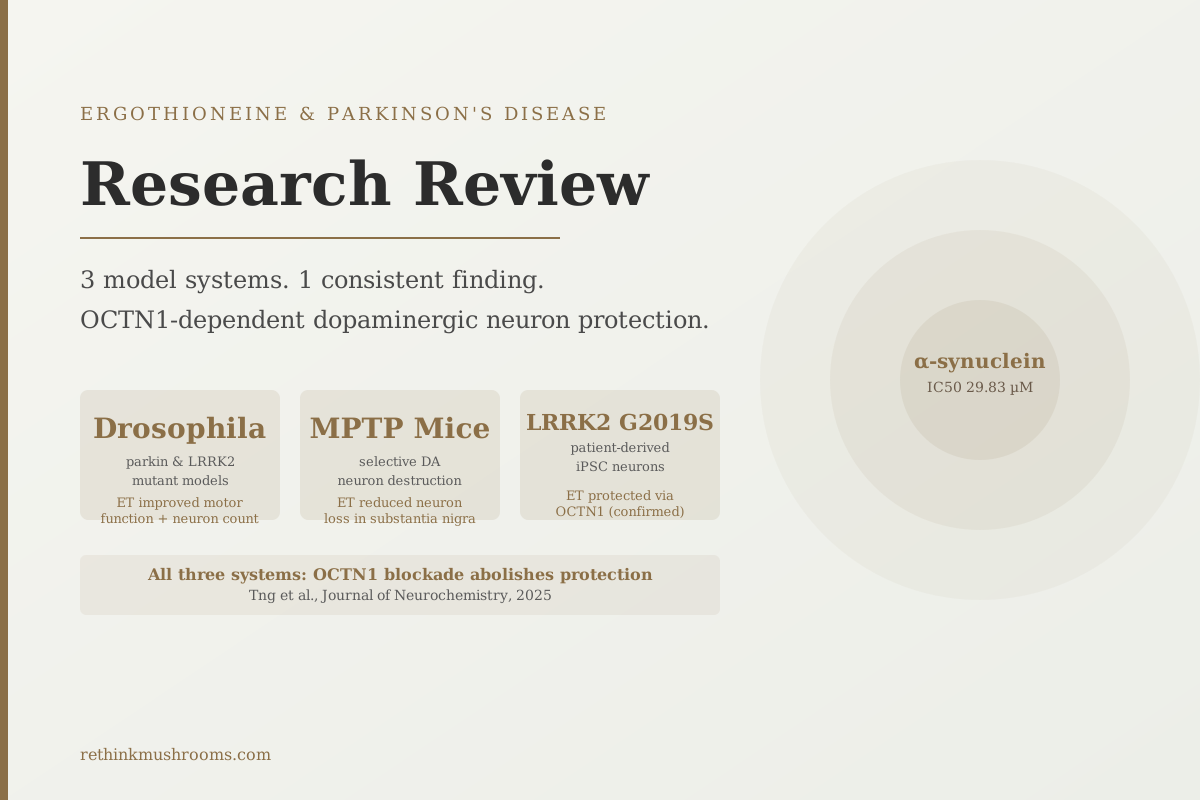
A 2025 Journal of Neurochemistry study tested ergothioneine across Drosophila mutants, MPTP mice, and patient-derived LRRK2 neurons simultaneously — the most comprehensive multi-model Parkinson's disease evidence published to date.
Tng et al., J Neurochem, 2025
OCTN1-dependent
protection confirmed: verapamil blocks all neuroprotective effects
29.83 µM IC50
concentration at which ET inhibits α-synuclein fiber formation
2016–2025
decade of Singapore research linking low ET to neurodegeneration
Tng et al., J Neurochem, 2025
 View as image ↗
Parkinson's disease destroys a specific population of neurons: the dopamine-producing cells in the substantia nigra that control movement. Whatever eventually kills them — mitochondrial failure, oxidative damage, toxic protein aggregates, or some combination of all three. Researchers have been searching for compounds that can interrupt that process. Ergothioneine, a mushroom-derived amino acid, has shown up consistently in that search: lower levels in PD patients, protective effects in dopaminergic cell models, and as of 2025, the most comprehensive multi-model PD evidence to date from a single research program.
This article reviews the research on ergothioneine and Parkinson's disease, from mechanism to model systems to the honest limits of what the data can support. For the epidemiological story from the Singapore research group that originated most of this work, see Ergothioneine and Parkinson's Disease: Epidemiological Evidence From Singapore. For a primer on how ergothioneine reaches the brain in the first place, see Ergothioneine — The Longevity Amino Acid.
Key Takeaways
- A 2025 Journal of Neurochemistry study found ergothioneine improved dopaminergic neuron survival and mitochondrial integrity across three independent Parkinson's models: Drosophila parkin/LRRK2 mutants, MPTP-treated mice, and patient-derived LRRK2 G2019S iPSC neurons (Tng et al., 2025)
- In 6-OHDA-treated human iPSC dopaminergic neurons, ergothioneine attenuated mitochondrial membrane potential loss, reduced mitochondrial ROS, and preserved ATP — all of which were abolished by verapamil (OCTN1 inhibitor), confirming the transporter-dependency (Leow et al., Antioxidants, 2024)
- Ergothioneine inhibits α-synuclein fiber formation, the protein aggregates central to Parkinson's pathology, with an IC50 of approximately 29.83 µM
- A 2024 hypothesis paper from the National University of Singapore proposed that age-related ergothioneine deficiency may be causally responsible for neurodegeneration including Parkinson's disease (Halliwell & Cheah, Free Radic Biol Med, 2024)
- In a Singapore elderly cohort of 600 people, blood ergothioneine was consistently lower in individuals with cognitive impairment, and the same group has documented lower ET in Parkinson's patients specifically (Cheah et al., 2016; Nakamichi et al., 2022)
- All evidence to date is preclinical or observational; no randomized controlled trial has tested ergothioneine in Parkinson's disease patients
How Ergothioneine Protects Dopaminergic Neurons: Five Mechanisms
Before walking through the evidence, it helps to understand what ergothioneine is doing inside a dopaminergic neuron. A 2022 Neurochemical Research review by Nakamichi and colleagues summarized the mechanistic landscape, and subsequent work has added granularity across five specific pathways:[4]
- α-Synuclein aggregation inhibition. Ergothioneine binds to α-synuclein, the protein that forms the toxic clumps (Lewy bodies) central to Parkinson's pathology, and inhibits fiber formation with an IC50 of approximately 29.83 µM. Lower concentrations than the drug target concentration required by many experimental α-synuclein inhibitors.
- Mitochondrial membrane potential preservation. Ergothioneine prevents the collapse of the voltage gradient across mitochondrial membranes that precedes neuronal death in dopaminergic cell models, keeping ATP production active under toxic stress.[2]
- Nrf2 and DJ-1 pathway activation. Ergothioneine activates the Nrf2 antioxidant response pathway and modulates DJ-1 (also known as PARK7), a protein whose loss-of-function mutations directly cause familial Parkinson's disease. DJ-1 normally protects against mitochondrial oxidative stress; ergothioneine appears to support this same protective function.[7]
- Neuroinflammation suppression. Ergothioneine reduces pro-inflammatory cytokine release from activated microglia and neurons, counteracting the neuroinflammatory environment that accelerates dopaminergic neuron loss in PD.[4]
- OCTN1-mediated intracellular delivery. Every neuroprotective effect documented to date requires ergothioneine to actually enter the cell via OCTN1. When researchers block OCTN1 with verapamil, all protective effects disappear, establishing that surface-level antioxidant activity is not the explanation — intracellular ergothioneine is required.[2]
For the full mechanistic account of how OCTN1 mediates ET's entry into brain cells specifically, see OCTN1 in the Brain: Why the Blood-Brain Barrier Lets Ergothioneine Through.
The Tng 2025 Study: Three PD Models, One Paper, the Strongest Evidence Yet
In 2025, Tng and colleagues — working within the Singapore research network that includes Cheah and Halliwell — published in the Journal of Neurochemistry what is now the most comprehensive multi-model Parkinson's disease study for ergothioneine.
The study tested ergothioneine across three independent experimental systems simultaneously:
Drosophila PD models. Using fruit flies with mutations in either parkin or LRRK2 genes — both of which cause familial Parkinson's disease in humans — ergothioneine treatment improved climbing scores (a motor function readout) and preserved the number of dopaminergic neurons and mitochondrial integrity in these models.
MPTP mouse model. Mice treated with MPTP, a neurotoxin that selectively destroys dopaminergic neurons and is the most widely used pharmacological PD model, showed reduced dopaminergic neuron loss in the substantia nigra when treated with ergothioneine.
LRRK2 G2019S patient-derived iPSC neurons. Neurons derived from stem cells taken from actual PD patients carrying the LRRK2 G2019S mutation — the most common genetic cause of familial Parkinson's disease — were exposed to rotenone, a mitochondrial toxin. Ergothioneine protected these cells from rotenone-induced toxicity via an OCTN1-dependent mechanism.[1]
The convergence across three systems at different levels of biological complexity (invertebrate, rodent, and human-derived cells) represents the most persuasive preclinical case for ergothioneine in Parkinson's disease published to date. The mechanistic finding — mitochondrial function preservation via OCTN1 uptake — was consistent across all three systems.
The inclusion of patient-derived cells matters specifically. Most cell experiments use cell lines or animal-derived neurons. LRRK2 G2019S iPSC neurons carry the actual genetic background of Parkinson's disease patients, making the protective effect directly relevant to a human genetic PD subtype rather than an approximated model.
Leow 2024: Human iPSC-Derived Dopaminergic Neurons and OCTN1 Dependency
Leow and colleagues published a separate iPSC-based study in Antioxidants in 2024, predating Tng 2025 and providing the first formal demonstration in human-derived dopaminergic neurons of ergothioneine's OCTN1-dependency.
The model used 6-OHDA (6-hydroxydopamine), a neurotoxin that simulates the selective dopaminergic cell death of Parkinson's disease, applied to human iPSC-derived dopaminergic neurons. Ergothioneine pre-treatment attenuated three specific cellular readouts:
- Mitochondrial membrane potential loss (the early signal of cellular energy collapse)
- Mitochondrial reactive oxygen species generation (oxidative damage from inside the cell)
- Loss of cellular ATP
Each of these protective effects was abolished when verapamil, an OCTN1 inhibitor, was added to the culture.[2]
This verapamil control is the methodological key. It rules out the possibility that ergothioneine is acting as an external antioxidant that scavenges ROS from the cell surface. The protection requires ergothioneine to get inside the cell via OCTN1, enter the mitochondria, and operate within the organelle where PD-related damage is occurring.
The Deficiency Hypothesis: PD as a Consequence of Running Low
In 2024, Halliwell and Cheah at the National University of Singapore published a hypothesis paper in Free Radical Biology and Medicine that reframed the entire field's question. Rather than asking "does ergothioneine protect against PD in a model," they asked: are age-related neurodegenerative diseases, including Parkinson's, caused by declining ergothioneine levels in the aging population?
Their argument rests on a chain: ergothioneine is not made by the body, it declines with age, it is lower in PD patients, animals without the OCTN1 transporter show no brain ergothioneine and accelerated neurological deterioration, and blocking OCTN1 abolishes every protective effect observed experimentally.[3]
This is a causal hypothesis, not a proven relationship. The observational data is consistent. The mechanistic data is supportive. What does not yet exist is a controlled prospective study showing that raising ergothioneine in humans reduces PD incidence or progression. The hypothesis motivates that trial; it does not complete it.
The 2025 systematic review by Takhor and Phan, covering 19 studies on ergothioneine and neurodegeneration, concluded that the antioxidative, anti-inflammatory, and antisenescence properties of ergothioneine slow age-related neurodegeneration markers across experimental systems, while noting that optimal clinical dosing and human interventional evidence for PD specifically remain gaps.[8]
Mitochondria: The Core Mechanism for Dopaminergic Vulnerability
Parkinson's disease has a specific relationship with mitochondrial dysfunction. Dopaminergic neurons in the substantia nigra are metabolically demanding — they fire at high rates and require exceptional energy throughput. Mitochondrial failure hits them disproportionately hard compared to other neuron types. Nearly every genetic PD mutation identified (parkin, LRRK2, DJ-1, PINK1) disrupts mitochondrial quality control in some way.
This is why the mitochondrial protection data from ergothioneine is particularly relevant to PD rather than just neurodegeneration generally.
Chen and colleagues demonstrated in 2024 in Food Function that ergothioneine supplementation in an aging mouse model restored hippocampal ATP levels and reduced mitochondrial reactive oxygen species. Pro-inflammatory cytokines IL-1β, TNF-α, and IL-6 all dropped measurably in ergothioneine-treated animals.[6] Although that study used an aging rather than a PD-specific model, the mitochondrial targets overlap directly with the pathways that make dopaminergic neurons selectively vulnerable in PD.
Ishimoto and Kato's 2022 FEBS Letters review confirmed that the midbrain — the brain region containing the dopaminergic neurons lost in Parkinson's — has among the highest OCTN1 expression levels in the brain. This means ergothioneine, when available in adequate blood supply, is actively concentrated in precisely the neurons most at risk in PD.[5]
For a detailed look at how ergothioneine protects mitochondria across tissues, see Ergothioneine and Mitochondrial Protection.
Paul 2022: The Broader Neuroprotective Framework
Paul's 2022 review in Antioxidants and Redox Signaling positioned ergothioneine as a stress vitamin, a compound that accumulates selectively in tissues under high oxidative burden, which is exactly the condition of dopaminergic neurons in PD brains.
The review documented ET's protective effects against NMDA excitotoxicity (excessive glutamate signaling that kills neurons), cisplatin-induced neurotoxicity (chemotherapy-related neural damage), and amyloid-β-induced learning and memory deficits in rodents. Nrf2 and BDNF pathway activation are both documented downstream of ergothioneine.[7]
The stress-vitamin framing matters for PD specifically: if ergothioneine preferentially concentrates in neurons under metabolic stress, its depletion with age would hit the most vulnerable neurons (the high-demand dopaminergic population) the hardest.
What Population Data Shows
Cheah and colleagues' 2016 Singapore cohort study found that plasma ergothioneine was significantly lower in people with cognitive impairment versus age-matched healthy controls, and declined with advancing age across 600 participants.[9] Nakamichi and colleagues' 2022 Neurochemical Research review confirmed that blood ergothioneine is consistently lower in Parkinson's disease patients relative to healthy subjects across the studies reviewed, placing PD alongside cognitive impairment and frailty as conditions characterized by ergothioneine deficiency.[4]
For the detailed narrative of the Singapore research program's decade-long tracking of this relationship, including how the group moved from population observations to laboratory mechanistic work, see Ergothioneine and Parkinson's Disease: Epidemiological Evidence From Singapore. For the parallel evidence on dementia prevention across population studies, see Ergothioneine and Dementia Prevention: Population Studies.
What the Evidence Cannot Yet Tell Us
Every piece of evidence reviewed here is either preclinical — cells, fruit flies, mice — or epidemiological: observing that PD patients have lower ergothioneine than healthy controls. Neither type of evidence is sufficient to conclude that supplementing ergothioneine prevents or treats Parkinson's disease in humans.
The Tng 2025 multi-model evidence is the most compelling mechanistic data yet published. The patient-derived LRRK2 G2019S neuron result is particularly striking because it uses cells with the actual genetic background of a human PD subtype. But "ergothioneine protected these cells from rotenone in a culture dish" does not equal "ergothioneine prevents Parkinson's disease progression in people."
The MCI clinical pilot from Singapore (14 subjects, 52 weeks, primarily a dementia prevention study) does not test PD outcomes. There is no registered PD-specific ergothioneine trial as of this writing. This is the largest gap in the evidence base: the mechanistic case is solid, the biological plausibility is strong, and the human interventional data for PD specifically is absent.
The Bottom Line
The 2025 Journal of Neurochemistry study from the Singapore research group tested ergothioneine across three independent Parkinson's disease model systems simultaneously, finding consistent dopaminergic neuron protection via mitochondrial preservation and OCTN1-dependent intracellular uptake. Earlier work with human iPSC-derived dopaminergic neurons confirmed that OCTN1 blockade abolishes protection, and ergothioneine inhibits α-synuclein aggregation with an IC50 of approximately 29.83 µM. Population data consistently find lower ergothioneine in PD patients. All evidence to date is preclinical or observational; no controlled human trial for Parkinson's disease exists and represents the primary evidentiary gap in this research area.
Frequently Asked Questions
Sources & References
- 1. Source details for this reference are not yet available.
- 2. Source details for this reference are not yet available.
- 3. Source details for this reference are not yet available.
- 4RTHM-01589·Nakamichi, Noritaka et al. (2022). “Ergothioneine and central nervous system diseases..” Neurochemical research.Study breakdown →PubMed →↩
- 5RTHM-01544·Ishimoto, Takahiro et al. (2022). “Ergothioneine in the brain..” FEBS letters.Study breakdown →PubMed →↩
- 6. Source details for this reference are not yet available.
- 7. Source details for this reference are not yet available.
- 8. Source details for this reference are not yet available.
- 9RTHM-00420·Cheah, Irwin K et al. (2016). “Ergothioneine levels in an elderly population decrease with age and incidence of cognitive decline; a risk factor for neurodegeneration?.” Biochemical and biophysical research communications.Study breakdown →PubMed →↩