Ergothioneine & Brain / Neurological Health
Zero brain ET without OCTN1
Mice bred without the OCTN1 gene have no measurable ergothioneine in their brains despite normal blood levels. There is no secondary route — OCTN1 is the only mechanism by which this dietary compound enters the brain.
Ishimoto & Kato, FEBS Lett, 2022
Cerebellum
highest OCTN1 expression in the brain — followed by midbrain and medulla
4 cell types
endothelial cells, neurons, neural stem cells, and microglia all express OCTN1
Verapamil
OCTN1 inhibitor that abolishes every documented neuroprotective effect of ET
Ishimoto & Kato, FEBS Lett, 2022
 View as image ↗
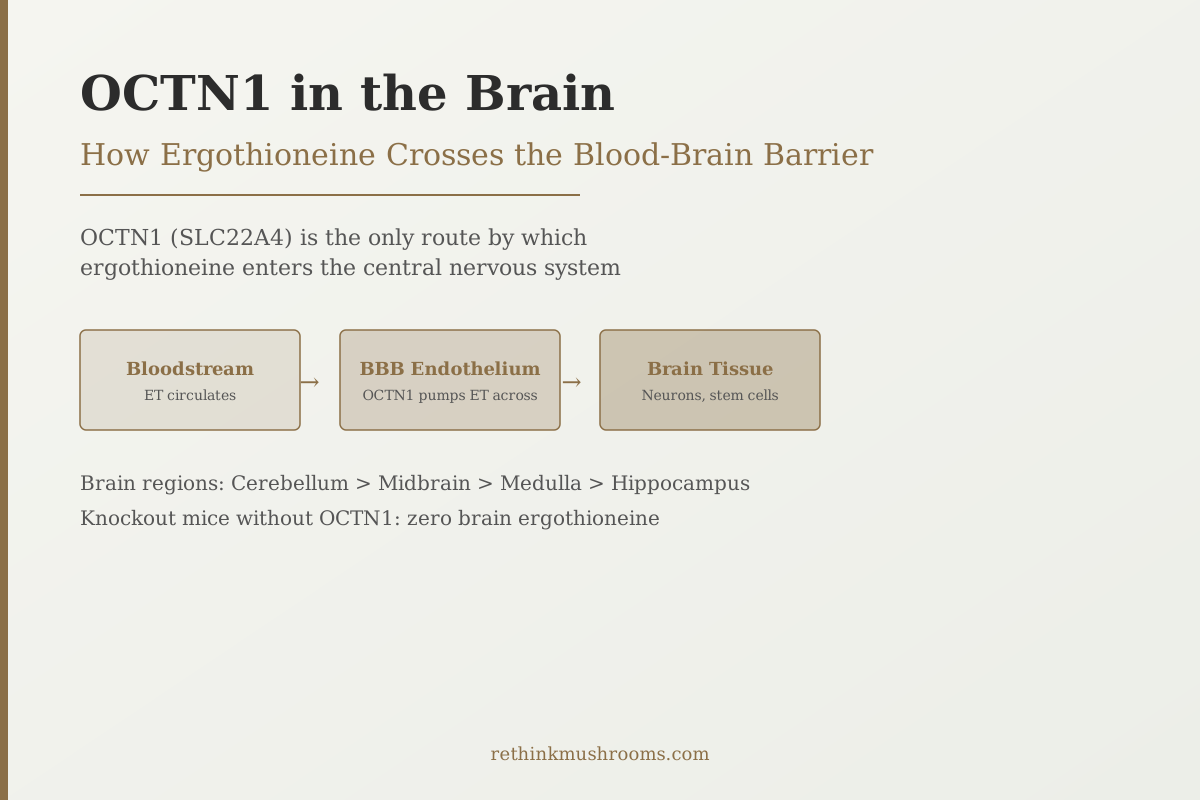
OCTN1 (gene: SLC22A4) is the dedicated ergothioneine transporter expressed in blood-brain barrier endothelial cells, neurons, neural stem cells, and microglia. It uses a proton gradient to pump ergothioneine across cell membranes. This is the only mechanism by which this dietary compound enters the brain — a fact confirmed by a single definitive experiment: mice without the OCTN1 gene have zero brain ergothioneine despite normal blood levels.
That exclusivity makes OCTN1 one of the more unusual facts in nutritional neuroscience. The blood-brain barrier exists to keep most molecules out of the central nervous system. Most dietary compounds either fail to cross it or slip through via nonspecific diffusion. Ergothioneine has a different arrangement: a dedicated molecular gate that actively pumps it across, concentrates it in specific brain regions, and has been encoded in the human genome for this purpose.
Ergothioneine is found almost exclusively in mushrooms and enters the body through diet. This article covers how it gets from the blood into the brain, where it goes once it's there, and why that distribution pattern matters for the neuroprotective effects documented in clinical and preclinical research.
Key Takeaways
- OCTN1 (SLC22A4) is the dedicated ergothioneine transporter expressed in blood-brain barrier endothelial cells — mice without OCTN1 have zero brain ET despite normal blood levels, proving it is the only entry route (Ishimoto & Kato, FEBS Lett, 2022)
- Gründemann's 2022 FEBS Letters inventory confirmed OCTN1's substrate specificity: it transports ET with high efficiency and selectivity, distinguishing it from general organic cation transporters
- OCTN1 expression is highest in the cerebellum, followed by midbrain, medulla, and hypothalamus — the midbrain distribution overlaps directly with the dopaminergic neurons lost in Parkinson's disease
- When OCTN1 is blocked with verapamil in cell experiments, every documented neuroprotective effect of ET disappears: mitochondrial protection, neuronal senescence clearance, and dopaminergic neuron protection all require ET to get inside cells via OCTN1
- Ishimoto et al. (2024) provided human functional proof: activated BDNF receptors in serum extracellular vesicles correlated with plasma ET in 52 volunteers, and TrkB inhibition in parallel mouse experiments abolished ET-driven neurogenesis — confirming the OCTN1-to-BDNF pathway operates in humans
- OCTN1 is expressed in neural progenitor cells and promotes neuronal differentiation — a finding that connects ET transporter expression to hippocampal neurogenesis independent of the antioxidant mechanism
What OCTN1 Is — and Why a Dietary Compound Having a Dedicated Transporter Is Unusual
OCTN1, encoded by the SLC22A4 gene, is a member of the organic cation transporter family. It sits in cell membranes and uses the proton gradient across those membranes to move molecules from one side to the other. Most members of this family move multiple substrates with varying efficiency. OCTN1 is different: it handles ergothioneine with exceptional selectivity and efficiency, which is precisely what makes ET's behavior unusual among dietary compounds.
Gründemann's 2022 FEBS Letters inventory of OCTN1 substrates and locations confirmed this specificity.[2] Most organic compounds that resemble ET structurally are transported poorly or not at all by OCTN1. ET, with its trimethylammonium group and thiol-imidazole structure, is the preferred substrate. This selectivity implies a functional relationship: the transporter appears to have been shaped by evolution specifically around ET rather than being a general-purpose mechanism that ET happens to fit.
Why does this matter? Because in nutrition, the existence of a dedicated transporter is taken as evidence that a compound serves a specific biological function. The body does not invest in a specific uptake mechanism for compounds it doesn't need. Vitamin C has a dedicated transporter. Folate has dedicated transporters. Ergothioneine is in this class — the only dietary amino acid derivative with a dedicated human uptake system.[2]
For the broader context of OCTN1 across all tissues (not just the brain), see OCTN1: The Dedicated Transporter That Proves Ergothioneine Matters.
How Ergothioneine Physically Crosses the Blood-Brain Barrier
The blood-brain barrier is a layer of endothelial cells — cells that line the interior of blood vessels — that forms an unusually tight seal around the brain's capillaries. Tight junction proteins connect these endothelial cells so closely that most molecules, including many drugs, cannot pass between them. Compounds can only cross by moving through the endothelial cells themselves, and the cells control what they absorb.
OCTN1 is expressed on the surface of these brain barrier endothelial cells. Ergothioneine circulating in the blood binds to OCTN1, which uses the proton gradient between the blood (slightly acidic) and the inside of the endothelial cell to actively pump ET across. This is an energy-requiring process, not passive diffusion — which is why it is directional and concentration-independent. Even when brain ET is already relatively high, OCTN1 continues to transport ET in, maintaining and concentrating it.
Ishimoto and Kato's 2022 FEBS Letters review documented this mechanism in detail, mapping OCTN1 expression at the blood-brain barrier and within brain tissue.[1] Once ergothioneine has crossed the endothelial wall, it encounters OCTN1 expressed on the neuronal cell surface, on neural stem cells, and on microglia (the brain's immune cells). Each of these cell populations uses its own OCTN1 to pull ET out of the extracellular space and concentrate it intracellularly.
The result is a layered accumulation system: blood to endothelium (first OCTN1), endothelium to extracellular space, extracellular space to neuron/stem cell/microglia (second OCTN1). This layered system produces the high intracellular ET concentrations required for the mitochondrial and anti-inflammatory effects documented in experimental systems.[1]
Syahputra and colleagues' 2025 mechanistic review confirmed that this intracellular accumulation specifically in high-stress environments is what drives ET's antioxidant specificity: OCTN1 is upregulated in cells under oxidative stress, meaning ET concentrates exactly where it is needed most.[9]
Which Brain Regions Have the Highest Ergothioneine: Cerebellum First
Ishimoto and Kato's 2022 review mapped OCTN1 mRNA expression and ET concentration across all major brain regions. The results were unexpected.[1]
The cerebellum had the highest OCTN1 expression of any brain region. The midbrain, medulla, and hypothalamus followed. The hippocampus showed lower but measurable OCTN1 expression. The cortex had the lowest expression of the regions measured.
This distribution is not what most people expect. The hippocampus, famous for its role in memory formation and its vulnerability in Alzheimer's disease, is not where ergothioneine concentrates most heavily. The cerebellum — which controls motor coordination and balance — and the midbrain are the primary targets of OCTN1-mediated ET delivery.
This distribution pattern has a direct implication for Parkinson's disease. The substantia nigra, the midbrain structure whose dopaminergic neurons are selectively destroyed in PD, sits within the midbrain region where OCTN1 expression is among the highest in the brain. These are precisely the neurons for which ET delivery is most robust — and precisely the neurons for which ET depletion with age would be most consequential. Nakamichi and colleagues' 2022 review confirmed this mechanistic connection between OCTN1 brain distribution and the selective vulnerability of PD-relevant neurons.[3]
For the full Parkinson's disease evidence, see Ergothioneine and Parkinson's Disease: Research Review.
The Knockout Experiment: No OCTN1 Means No Brain Ergothioneine
The most definitive evidence that OCTN1 is the exclusive brain entry route for ergothioneine comes from a genetic experiment: removing the OCTN1 gene entirely from mice and measuring what happens.
Mice bred without a functional OCTN1 gene (OCTN1 knockout mice) show normal plasma ergothioneine levels. The gut absorbs ET normally because other transporters contribute to intestinal uptake. But when researchers measure brain tissue in these knockout animals, the result is absolute: zero detectable ergothioneine.[1]
This is not a reduction — it is complete absence. Despite circulating blood ET at normal concentrations, not a single molecule reaches the brain without OCTN1.
The experiment eliminates any possibility that ET crosses the blood-brain barrier through nonspecific diffusion, through lipid partitioning (the mechanism by which fat-soluble compounds cross), or through any other transporter. The brain compartment is sealed against ET in the absence of OCTN1. This level of exclusivity is not observed for most nutrients; it places ergothioneine's brain delivery on an entirely different mechanistic footing from, say, vitamin E or omega-3 fatty acids, which distribute throughout the brain via lipid-based mechanisms.
The Gründemann 2022 inventory confirmed the same conclusion from the transporter biology perspective, finding no evidence of a secondary transport mechanism that could substitute for OCTN1's ET-delivery function.[2]
What Happens When You Block OCTN1: The Verapamil Experiments
The knockout genetic experiment showed that removing OCTN1 eliminates brain ET. A complementary pharmacological approach uses verapamil, a drug known to inhibit OCTN1 function, to block the transporter in cells that would otherwise express it normally. The results of these experiments reveal exactly which of ET's effects require OCTN1-mediated intracellular delivery.
Three studies from 2024 independently applied this test:
Apparoo et al. (2024) tested ET's ability to clear senescent neurons — cells that have stopped working but remain active and emit inflammatory signals. Ergothioneine-rich mushroom extract selectively eliminated these senescent neurons. When OCTN1 was blocked with verapamil, the clearance stopped completely. ET had no effect on senescent neurons when it couldn't get inside them.[5]
Leow et al. (2024) used human iPSC-derived dopaminergic neurons modeled on Parkinson's disease. Ergothioneine protected these neurons from 6-OHDA-induced mitochondrial damage: it preserved mitochondrial membrane potential, reduced mitochondrial ROS, and maintained ATP. Adding verapamil to block OCTN1 abolished all three protective effects.[7]
Tng et al. (2025) extended this OCTN1-dependency confirmation across three independent Parkinson's disease model systems simultaneously. In Drosophila PD mutants, MPTP-treated mice, and LRRK2 G2019S patient-derived neurons, the mitochondrial protection ET provided was mechanistically linked to OCTN1-mediated intracellular uptake.[8]
The conclusion from these parallel experiments is consistent: ergothioneine's neuroprotective effects are entirely dependent on OCTN1. Surface-level antioxidant activity, if any exists, is insufficient. ET must physically enter the cell through OCTN1 to protect it.
This specificity has clinical implications. A compound whose effects are transporter-dependent is not simply "more antioxidant." It is a compound whose delivery, distribution, and action are regulated by a specific biological mechanism — the same mechanism that age-related depletion would compromise.
OCTN1 in Neural Stem Cells: The Neurogenesis Connection
Beyond its role in mature neurons and microglia, OCTN1 is expressed in neural progenitor cells — the stem cells in the hippocampus that generate new neurons throughout adult life. ET delivered via OCTN1 to these cells appears to promote neuronal differentiation rather than just protecting existing neurons.
Ishimoto and colleagues demonstrated this in their 2024 study in NPJ Science of Food. In 52 volunteers taking 5 mg ET daily for 12 weeks, activated TrkB receptors measured in serum extracellular vesicles were significantly higher than placebo and correlated with both plasma ET levels and cognitive test scores.[4]
In parallel mouse experiments, ET-deficient animals showed reduced hippocampal neurogenesis. Supplementing ET restored new neuron formation. Blocking TrkB pharmacologically abolished the neurogenesis rescue — confirming the pathway was BDNF/TrkB dependent. And since OCTN1 is required for ET to reach neural progenitor cells in the first place, the entire neurogenesis mechanism is OCTN1-dependent at its first step.
Chen and colleagues' 2024 Food Function study added the metabolic dimension: in aging mice, ET supplementation improved hippocampal mitochondrial membrane potential and reduced neuroinflammatory cytokines IL-1β, TNF-α, and IL-6. These improvements, again, require ET delivery via OCTN1 into hippocampal cells.[6]
For the complete picture of ET's neuroprotective mechanisms including BDNF, senescent neuron clearance, and neuroinflammation reduction, see Ergothioneine and Brain Health: Neuroprotective Research.
For the population evidence connecting brain ET levels to dementia risk, see Ergothioneine and Dementia Prevention: Population Studies.
For the full Singapore epidemiological story behind this research, see Ergothioneine and Parkinson's Disease: Epidemiological Evidence From Singapore.
For a compare/contrast between ET's OCTN1-dependent BBB crossing and Lion's Mane's erinacines, which cross via a different mechanism, see Erinacines: The Compounds That Cross the Blood-Brain Barrier.
The Bottom Line
OCTN1 (SLC22A4) is the exclusive route by which ergothioneine enters the brain. It is expressed in blood-brain barrier endothelial cells, neurons, neural stem cells, and microglia, and uses a proton gradient to actively pump ET into each cell type. The cerebellum and midbrain show the highest OCTN1 expression — regions directly relevant to Parkinson's disease vulnerability. OCTN1 knockout mice have zero brain ET. Pharmacological OCTN1 blockade (verapamil) abolishes every documented neuroprotective effect of ET across three independent 2024 studies, confirming that intracellular delivery is required for all observed protection. Human functional evidence shows OCTN1-mediated brain ET activates BDNF/TrkB-driven neurogenesis at 5 mg/day over 12 weeks.
Frequently Asked Questions
Sources & References
- 1RTHM-01544·Ishimoto, Takahiro et al. (2022). “Ergothioneine in the brain..” FEBS letters.Study breakdown →PubMed →↩
- 2RTHM-01527·Gründemann, Dirk et al. (2022). “The ergothioneine transporter (ETT): substrates and locations, an inventory..” FEBS letters.Study breakdown →PubMed →↩
- 3RTHM-01589·Nakamichi, Noritaka et al. (2022). “Ergothioneine and central nervous system diseases..” Neurochemical research.Study breakdown →PubMed →↩
- 4. Source details for this reference are not yet available.
- 5. Source details for this reference are not yet available.
- 6. Source details for this reference are not yet available.
- 7. Source details for this reference are not yet available.
- 8. Source details for this reference are not yet available.
- 9. Source details for this reference are not yet available.