Lion's Mane Bioactive Compounds: Hericenones & Erinacines
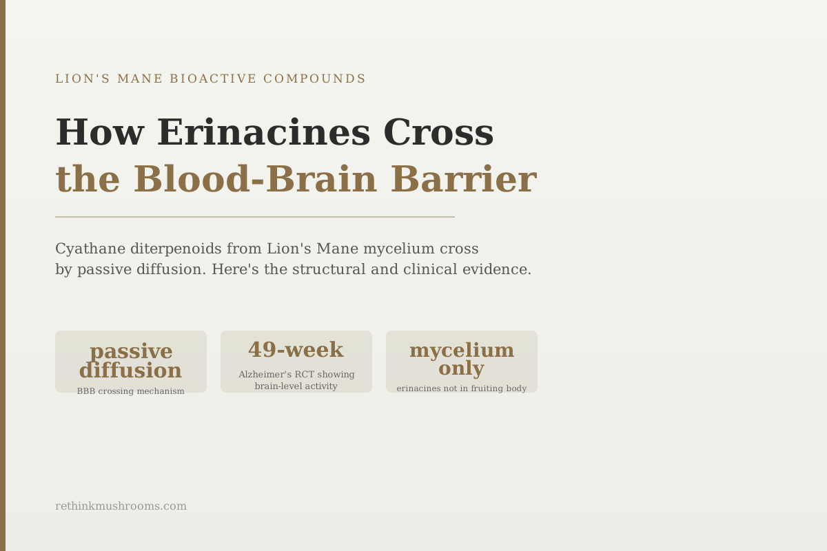
49-week RCT
In a 49-week placebo-controlled trial, erinacine A-enriched Lion's Mane improved daily function and reduced amyloid deposits in early Alzheimer's patients, demonstrating that brain-penetrating erinacines produce measurable outcomes.
Lee et al., Frontiers in Aging Neuroscience, 2020
passive diffusion
the mechanism by which erinacines cross the BBB; no active transport is required; driven by their fat-soluble diterpenoid structure
mycelium only
the part of Lion's Mane that contains erinacines; fruiting body products contain none (Chen 2017 genomics)
2024
erinacine S confirmed neuroprotective in ischemic brain injury model, expanding the family's brain evidence base
Lee et al., Frontiers in Aging Neuroscience, 2020
 View as image ↗
Most compounds that reach the bloodstream never reach the brain. The blood-brain barrier, a tightly sealed membrane of specialized cells lining the brain's blood vessels, blocks nearly everything from crossing into neural tissue. Small molecules, toxins, pathogens, and most drugs are filtered out. To enter the brain, a compound either needs to use one of the specific transport proteins embedded in the barrier or be physically small and fat-soluble enough to dissolve through the barrier's fat-based membrane on its own.
Erinacines, the diterpenoid compounds found exclusively in Lion's Mane mycelium, belong to the second category. Their molecular architecture makes them naturally equipped for this crossing. This article covers how that works structurally, what the evidence shows about what they do once inside the brain, and why the mycelium-exclusive nature of these compounds matters for supplement selection. For the broader overview of erinacine and hericenone chemistry, see hericenones and erinacines: Lion's Mane's brain-boosting compounds and the bioactive compounds in mushrooms pillar.
Key Takeaways
- Erinacines cross the blood-brain barrier via passive diffusion: their fat-soluble cyathane diterpenoid structure allows them to dissolve through the lipid membrane of the barrier without needing active transport
- Erinacines are exclusively produced in the mycelium due to CYP450 enzyme gene clusters that are expressed in mycelium but not in fruiting bodies; Lion's Mane fruiting body products contain no erinacines
- A 2018 study confirmed erinacine A activates the BDNF/PI3K/Akt/GSK-3β pathway in the hippocampus after oral administration, demonstrating functional brain activity following the crossing
- A 49-week placebo-controlled trial in early Alzheimer's patients found erinacine A-enriched mycelium improved daily living scores and reduced amyloid deposit markers, providing human-level evidence of brain-reaching activity
- A 2021 Scientific Reports study found Lion's Mane mycelium promotes oligodendrocyte maturation and myelin basic protein expression, a second BBB-enabled mechanism distinct from NGF stimulation
- Erinacine S, a related compound, was confirmed neuroprotective in ischemic brain injury (2024), expanding the evidence that multiple erinacine variants cross the barrier and act in brain tissue
What the Blood-Brain Barrier Is and Why Most Compounds Fail to Cross It
The blood-brain barrier is not a wall. It is a continuous layer of specialized cells called endothelial cells that line every blood vessel feeding the brain and spinal cord. Unlike the blood vessel cells elsewhere in the body, these endothelial cells are sealed together with very tight junctions, leaving almost no gaps. This prevents almost everything dissolved in the blood from leaking into brain tissue.
The brain can still get the nutrients it needs because it has specific transport proteins embedded in these cells. Glucose has a transporter. Amino acids have transporters. Essential nutrients are pulled through selectively. But most drugs, supplements, and bioactive compounds don't have dedicated transporters. They need to cross by dissolving through the cell membrane itself.
Cell membranes are made primarily of phospholipids, which are fat-based molecules. For a compound to dissolve through a membrane, it needs to be fat-soluble (the technical term is lipophilic). Water-soluble compounds get blocked at the membrane surface and cannot cross. The degree of fat-solubility a compound needs to passively cross the blood-brain barrier is specific: too little and it can't dissolve in; too much and it gets stuck in the membrane without passing through.
Small, moderately fat-soluble molecules with low molecular weight tend to cross. This is why caffeine, ethanol, and many small drug molecules reach the brain quickly. Larger, water-soluble compounds like most polysaccharides, proteins, and vitamin B12 do not cross without specialized carriers.
The Cyathane Skeleton: Why Erinacines Are Structurally Built for Brain Entry
Erinacines are cyathane diterpenoids. The word "cyathane" refers to a specific molecular architecture: three rings fused together (containing 5, 6, and 7 carbons respectively), creating the backbone around which all erinacine compounds are built. This fused-ring structure, combined with attached oxygen-containing groups, gives erinacines a fat-soluble character while keeping their molecular weight low enough for passive membrane diffusion.
A 2018 comprehensive review of erinacine research by Roda and colleagues confirmed that erinacine A crosses the blood-brain barrier based on cumulative pharmacological and functional evidence.[1] The review catalogued all known erinacines (A through I at that time) and their documented biological activities, noting that erinacine A is the most studied and has the clearest evidence for brain penetration.
The genomic basis for erinacines' exclusive occurrence in mycelium was established by Chen and colleagues in a 2017 Scientific Reports study analyzing the Hericium erinaceus genome.[2] The researchers identified three distinct terpene biosynthesis gene clusters in the mushroom's genome, along with a large family of CYP450 enzymes responsible for constructing the cyathane diterpenoid skeleton step by step. These gene clusters are expressed in mycelium but not in fruiting bodies, which is why erinacines simply do not form in the part of the mushroom people recognize as the cap and stem. A Lion's Mane fruiting body extract, regardless of quality, contains no erinacines.
This distinction matters practically. The brain-penetrating compounds in Lion's Mane require a mycelium-sourced product. For readers exploring this supplement category, erinacines: the compounds that cross the blood-brain barrier covers the compound profile in more depth, and isolation and characterization of Lion's Mane diterpenoids covers the structural chemistry.
Functional Proof: What Erinacines Do Once Inside the Brain
The clearest evidence that erinacines cross the blood-brain barrier isn't a direct measurement of erinacine concentration in brain tissue. It is functional: studies show erinacines activating specific brain pathways that can only be activated from inside neural tissue.
Chiu and colleagues published a study in the International Journal of Molecular Sciences in 2018 testing erinacine A-enriched Hericium erinaceus mycelium in mice.[3] After oral administration, mice showed activation of the BDNF/PI3K/Akt/GSK-3β signaling pathway in the hippocampus (the brain's primary memory region). In plain terms: a chain of signaling proteins that tells neurons to survive, grow, and adapt was turned on in a brain region, after the compound was swallowed. This cascade can only be activated by something that has physically entered the brain.
The endpoint was antidepressant-like behavior: the erinacine A group showed reductions in behavioral markers of depression and enhanced hippocampal neurogenesis (new neuron growth) compared to controls.[3]
Lai and colleagues showed in a 2013 in vitro study that when Lion's Mane extract combined with nerve growth factor was applied to neural cells, neurite outgrowth (the growth of nerve fiber extensions) increased by 60.6% compared to nerve growth factor alone, with the signaling running through MEK/ERK and PI3K/Akt pathways.[9] The downstream signaling these pathways activate in hippocampal tissue is the same circuitry the Chiu 2018 study documented in living animals.
The combined picture: erinacines reach the brain following oral administration, then activate well-characterized neural signaling pathways with measurable behavioral and histological consequences.
The TBI Finding: Evidence That Brain Penetration Is Fast Enough to Matter Therapeutically
A 2020 study by Lee and colleagues published in the International Journal of Molecular Sciences tested erinacine A after traumatic brain injury in an animal model.[4] The design was post-treatment: erinacine A was administered after the injury occurred, not prophylactically beforehand. The compound reduced cerebral infarct volume (the amount of brain tissue killed) and improved neurological outcomes.
The therapeutic relevance of a post-treatment design is that it implies the compound crossed the blood-brain barrier rapidly enough to be present in brain tissue at the time of acute injury response. Compounds that take days to cross and accumulate are less relevant to acute neuroprotection. Erinacine A's documented efficacy in post-injury TBI settings is consistent with pharmacokinetic evidence that it reaches brain tissue within a pharmacologically relevant timeframe after administration.
The Alzheimer's Trial: Human Evidence of Brain-Level Activity
The most clinically significant evidence for erinacine A's brain-penetrating activity comes from a human trial. Lee and colleagues published a 49-week, double-blind, placebo-controlled pilot study in Frontiers in Aging Neuroscience in 2020, testing erinacine A-enriched Hericium erinaceus mycelium in patients with early Alzheimer's disease.[5]
The erinacine A group showed improved activities of daily living (ADL) scores and, on cognitive testing, improved MMSE (Mini-Mental State Examination) scores compared to placebo. They also showed reduced amyloid-β deposition markers (amyloid-β is the sticky protein that accumulates in Alzheimer's brain tissue as plaques).
This trial is a pilot study with a small sample size and early-phase evidence, not definitive. But it represents the strongest human evidence available that erinacine A produces changes in brain tissue in living people. Amyloid-β reduction in the brain requires a compound to physically reach neural tissue, interact with the amyloid-β pathway, and produce measurable changes over weeks. Erinacine A meeting that bar in a controlled trial is the most direct available evidence for functional brain penetration.
For the broader Alzheimer's and cross-species mushroom context, see cross-species mushroom research in Alzheimer's: Lion's Mane, Reishi, and Cordyceps.
A Second Brain Mechanism: Oligodendrocyte Maturation and Myelin Repair
Most coverage of erinacines in the brain focuses on NGF stimulation and its downstream cognitive effects. A 2021 study by Ryu and colleagues in Scientific Reports documented a second, distinct mechanism that also requires brain entry: effects on oligodendrocytes, the cells that produce the myelin sheath wrapping nerve fibers.[6]
Lion's Mane mycelium extracts promoted oligodendrocyte maturation and increased myelin basic protein (MBP) expression, the structural protein that forms part of the myelin sheath. Because oligodendrocytes are in the brain and spinal cord, these changes can only occur if the relevant compounds have crossed the blood-brain barrier and reached neural tissue.
This myelin-supporting mechanism is separate from the NGF pathway. It suggests erinacines have at least two distinct modes of action in the brain: one that drives neurotrophic signaling through NGF and BDNF pathways, and one that supports the physical structure of the neurons' insulating coating. Both require successful BBB penetration.[6]
Beyond NGF: The Pan-Neurotrophic ERK1/2 Discovery
The established model of how erinacines and hericenones support brain health has been: compounds from Lion's Mane stimulate cells to produce nerve growth factor (NGF), which then signals neurons to grow and maintain their connections. This model is accurate but incomplete.
Martínez-Mármol and colleagues published a study in the Journal of Neurochemistry in 2023 finding that hericerin derivatives (compounds structurally related to hericenones) activate a separate neurotrophic signaling pathway in hippocampal neurons that operates independently of NGF protein.[7] The pathway runs through ERK1/2, a signaling molecule that several different growth factors all converge on. When activated, it tells neurons to grow, form connections, and survive. Mice fed hericene A showed enhanced spatial memory performance.
This is called a "pan-neurotrophic" effect because ERK1/2 sits downstream of multiple growth factor receptors. Activating it directly bypasses the NGF secretion step entirely. The finding updates the mechanism model in a practically significant way: the neuroactive effects of Lion's Mane compounds are not limited to promoting NGF secretion; some compounds activate neurotrophic signaling directly in hippocampal neurons.[7]
Erinacine S and the Expanding Compound Family
Erinacine A has received the most research attention, but the erinacine family now includes 15 or more identified compounds, several of which have their own documented brain-related activity.
A 2018 review by Roda and colleagues documented that erinacine E reduces neuropathic pain through central mechanisms and erinacine S showed stroke protection in prior animal models.[1]
A 2024 study by Chen and colleagues specifically tested erinacine S in a model of ischemic brain injury, finding neuroprotection, confirming that this structurally distinct erinacine variant also reaches and acts in brain tissue.[8]
The picture that emerges is of a structurally diverse family of brain-penetrating compounds, each with specific therapeutic profiles, all dependent on mycelium sourcing for their production. For the broader NGF context, see Lion's Mane and nerve growth factor: the complete research guide. For the hericenone side of Lion's Mane (the NGF-stimulating compounds exclusive to the fruiting body), see hericenone C and NGF synthesis: the key mechanism and hericenones A-H: mapping the full library.
The Bottom Line
Erinacines cross the blood-brain barrier via passive diffusion enabled by their fat-soluble cyathane diterpenoid structure. The genomic reason they occur only in mycelium (not fruiting body) is the expression of CYP450 biosynthesis gene clusters in mycelium tissue only. Functional evidence of brain penetration includes hippocampal BDNF/PI3K/Akt pathway activation after oral administration (Chiu 2018), post-traumatic brain injury protection after treatment (Lee 2020), and improved brain outcomes in a 49-week Alzheimer's RCT (Lee 2020). A second BBB-enabled mechanism involves oligodendrocyte maturation and myelin protein expression (Ryu 2021). A pan-neurotrophic ERK1/2 pathway discovered in 2023 shows that erinacine-related compounds activate neurotrophic signaling in hippocampal neurons independently of NGF protein secretion.
Frequently Asked Questions
Sources & References
- 1. Source details for this reference are not yet available.
- 2. Source details for this reference are not yet available.
- 3. Source details for this reference are not yet available.
- 4. Source details for this reference are not yet available.
- 5. Source details for this reference are not yet available.
- 6. Source details for this reference are not yet available.
- 7. Source details for this reference are not yet available.
- 8. Source details for this reference are not yet available.
- 9. Source details for this reference are not yet available.