Meshima & Immune System
4 DC maturation markers upregulated
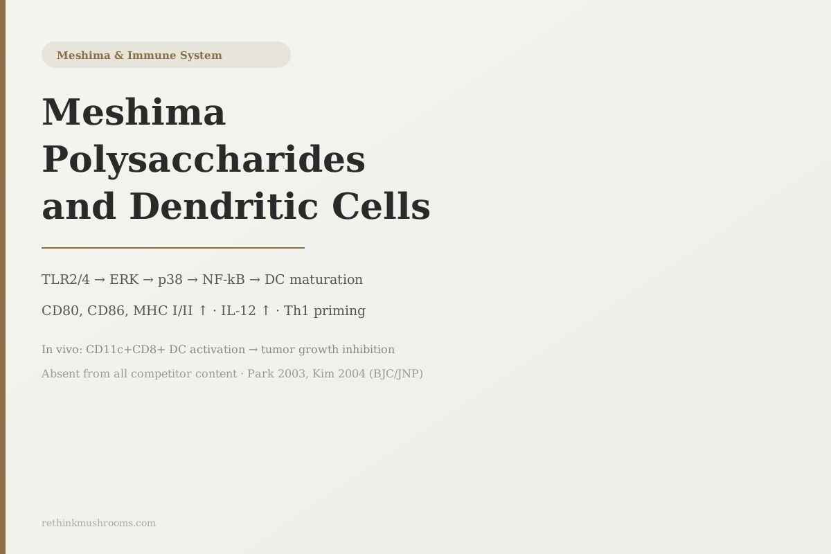
PL-treated dendritic cells showed increased CD80, CD86, MHC I, and MHC II expression, secreted higher IL-12, and migrated preferentially to lymphoid tissues, making them more potent antigen presenters.
Park et al., J Nat Prod, 2003
TLR2 + TLR4
both pattern-recognition receptors activated by PL proteoglycan (Kim 2004)
CD11c+CD8+
specialized dendritic cell subset activated in vivo, driving Th1 anti-tumor immunity (Kim 2004)
IL-12
cytokine that switches T cells toward Th1 responses, significantly elevated in PL-treated DCs (Park 2003)
Park et al., J Nat Prod, 2003
 View as image ↗
Dendritic cells are the immune system's briefing agents. They patrol body tissues, sample antigens from pathogens or damaged cells, mature into a fully activated state, and then travel to lymph nodes to present those antigens to T cells. That briefing is what launches the adaptive immune system: the slow, precise arm of immunity that builds targeted memory against specific threats. Without dendritic cell maturation, T cells have no information to act on.
Three published studies from 2003 and 2004, all in peer-reviewed journals, established that specific polysaccharide fractions from Phellinus linteus (meshima) trigger dendritic cell maturation through a measurable molecular pathway. The findings are unusually specific: they name the receptors involved, the signaling cascade that follows, the surface proteins that change on the DCs, the cytokines those DCs produce, and the downstream T cell response the whole cascade generates. No consumer content on meshima covers any of this.
This article works through that cascade from beginning to end. For the foundational introduction to meshima, see What Is Meshima Mushroom. For the underlying compounds responsible for these effects, see Phellinus linteus Bioactive Compounds.
Key Takeaways
- Park et al. (2003, Journal of Natural Products) found that PL acidic polysaccharides induced dendritic cell maturation: CD80, CD86, MHC I, and MHC II all increased on the cell surface, IL-12 secretion was elevated, and treated DCs migrated preferentially to lymphoid tissues where T cells wait
- Kim et al. (2004, Biological and Pharmaceutical Bulletin) identified the receptor pathway: PL proteoglycan binds TLR2 and TLR4, activating ERK, p38, and NF-κB cascades that drive dose-dependent increases in MHC II, CD86, and IL-12
- Kim et al. (2004, Journal of Natural Products) confirmed the downstream outcome in vivo: PL proteoglycan inhibited tumor growth in mice by activating CD11c+CD8+ dendritic cells and driving a Th1-dominant immune state with elevated IL-12 and IFN-gamma
- The same acidic polysaccharide fractions also activated macrophages through PTK/PKC pathways, producing tumoricidal nitric oxide: DC and macrophage activation appear to be parallel tracks from the same PL compound class (Kim 2003, 2004)
- A 2023 systematic review by Qin et al. confirmed that immunomodulatory potency depends critically on polysaccharide structure: branching patterns and molecular weight determine the strength of TLR signaling and downstream immune activation, explaining why commercial extracts vary in potency
- All mechanistic DC data is preclinical (in vitro and mouse models); the two 2022 Korean RCTs confirmed human NK cell improvements from PL supplementation, but did not directly measure dendritic cell activation
What Dendritic Cells Do and Why Activation Markers Matter
Dendritic cells exist in two functional states: immature and mature. Immature DCs patrol tissues and take up antigens efficiently, but they cannot effectively prime T cells. They are collectors, not communicators. The transition from immature to mature DC is what makes the difference. Mature DCs express a specific set of surface proteins that T cells need to recognize before responding: the co-stimulatory molecules CD80 and CD86, and the antigen-presentation proteins MHC class I and MHC class II. Without these, a T cell that encounters an antigen either ignores it or becomes anergic (permanently unresponsive).
CD80 and CD86 are the "permission" signals. They bind to CD28 on T cells, providing the second signal that allows T cell activation to proceed. An antigen presented without CD80/CD86 signaling leads to tolerance rather than immunity. MHC class II presents antigens to CD4+ helper T cells; MHC class I presents to CD8+ cytotoxic T cells. Upregulation of both means a mature DC can prime both arms of the T cell response.
IL-12 is the cytokine that determines which direction the T cell response goes. High IL-12 from DCs pushes CD4+ T cells toward a Th1 response: interferon-gamma (IFN-gamma) production, cytotoxic T cell activation, and macrophage activation. Th1 responses are the primary defense against intracellular pathogens and cancer cells. Low IL-12 pushes toward Th2: antibody production, allergy, and parasitic defense.
A compound that drives DC maturation (CD80, CD86, MHC up) and increases IL-12 production is therefore a compound that biases the whole adaptive immune response toward the Th1 arm. That is the significance of the Park 2003 finding.
The Park 2003 Discovery: What PL Polysaccharides Do to Immature DCs
Park et al. (2003) isolated acidic polysaccharides from Phellinus linteus and exposed immature murine dendritic cells to them, measuring the resulting phenotypic and functional changes.[1] The results covered each component of DC maturation.
Surface phenotype: CD80, CD86, MHC class I, and MHC class II all increased significantly on PL-treated DCs compared to untreated controls. All four are markers of mature, fully functional antigen-presenting cells. The treated DCs looked, phenotypically, like DCs that had just encountered a pathogen.
Functional capacity: PL-treated DCs were more potent antigen-presenting cells when tested in mixed leukocyte reactions (a standard assay for DC-T cell interaction quality). They provoked stronger T cell responses than untreated DCs when both were loaded with the same antigen.
IL-12 secretion: DCs treated with PL polysaccharides produced significantly more IL-12 than untreated DCs. As described above, this is the cytokine that drives the Th1 response.
Migration behavior: PL-stimulated DCs migrated preferentially toward lymphoid tissues in chemotaxis assays. This is mechanistically significant: a DC that has collected antigen needs to reach a lymph node to brief T cells. A DC that migrates toward lymph nodes more readily is more likely to complete that briefing before the antigen degrades.
The Receptor Pathway: How TLR2 and TLR4 Are Activated
Park 2003 established what happens to DCs when they encounter PL polysaccharides. Kim et al. (2004) established how it happens at the molecular level.[2]
TLR2 and TLR4 (toll-like receptors 2 and 4) are pattern recognition receptors on the surface of immune cells. They evolved to detect conserved molecular patterns from pathogens: TLR4 primarily detects bacterial lipopolysaccharide (LPS); TLR2 detects bacterial peptidoglycans and lipoproteins. When activated, both trigger intracellular signaling cascades that lead to inflammatory cytokine production and immune cell activation.
Kim et al. isolated a proteoglycan fraction from Phellinus linteus and showed it induced dose-dependent increases in MHC II and CD86 surface expression on DCs, stimulated IL-12 production, and enhanced T cell proliferation in allogeneic reactions. Critically, they identified the signaling pathway: TLR2/4 binding → ERK activation → p38 activation → NF-κB nuclear translocation. Blocking TLR2/4 with antibodies or using signaling inhibitors reduced DC activation. The pathway was not just observed; it was causally validated.
The mechanistic parallel to bacterial pattern recognition is notable. PL proteoglycan appears to trigger DC maturation through the same receptors that would detect bacterial infection, essentially signaling "danger" to the immune system. This may explain why DCs respond so robustly to the compound: they are using pattern recognition machinery that evolved for detecting genuine threats, and PL's proteoglycan fits the molecular template.
For how this NF-κB activation pathway also connects to meshima's anti-inflammatory properties in a different immune context, see Meshima and Inflammation: NF-kB Pathway Research.
From DCs to Anti-Tumor Immunity: The in Vivo Confirmation
Park 2003 and Kim 2004 (Biol Pharm Bull) established the cellular and molecular events in vitro. A second Kim et al. (2004) study, published in the Journal of Natural Products, confirmed the outcome in a living organism.[3]
In tumor-bearing mice, PL proteoglycan administration significantly inhibited tumor growth compared to controls. The study identified the immune mechanism: activation of CD11c+CD8+ dendritic cells. CD11c+CD8+ DCs are a specialized subset involved in cross-presentation, the process by which DCs present antigens from cancer cells to cytotoxic CD8+ T cells. This is the DC subset most directly involved in anti-tumor immunity.
The treated mice showed elevated IL-12 and IFN-gamma in tumor-associated tissue. These are the defining cytokines of a Th1-dominant immune state. The immune response had shifted from a neutral or Th2-dominant background toward the Th1 arm, and tumor growth was suppressed.
The chain from compound to outcome: PL proteoglycan binds TLR2/4 on DCs → DCs mature and express CD80/CD86/MHC/IL-12 → activated DCs (especially CD11c+CD8+) prime cytotoxic T cells → IL-12/IFN-gamma production confirms Th1 shift → tumor growth inhibited.
Macrophages: The Parallel Track
The studies above focus on dendritic cells. Two studies by Kim et al. establish that the same acidic polysaccharide fractions also activate macrophages through a distinct but parallel pathway.
Kim et al. (2003) found that PL acidic polysaccharide activated tumoricidal nitric oxide production in macrophages through protein tyrosine kinase (PTK) and protein kinase C (PKC) signaling cascades.[4] NO-mediated macrophage tumoricidal activity is one of the primary ways activated macrophages kill cancer cells. Kim et al. (2004) extended this to primary peritoneal macrophages (cells isolated directly from the mouse peritoneum, not cell lines), showing enhanced NO and TNF-alpha production.[5]
The significance of the parallel tracks: DCs and macrophages are both innate immune cells that PL polysaccharides activate. DCs bridge innate to adaptive immunity (briefing T cells). Macrophages act as direct killers. Both are activated by the same compound class. For a complete look at how meshima coordinates innate and adaptive immune activation, see How Meshima Bridges Innate and Adaptive Immunity.
Compound Class and Structural Specificity
Not all Phellinus linteus polysaccharides activate DCs equally. The immunological activity depends on specific structural features.
Kim et al. (2006) characterized a novel polysaccharide-protein complex from PL that showed immunostimulatory effects, establishing that the immune-active compounds include protein-polysaccharide conjugates, not just free polysaccharides.[6] The proteoglycans in the Kim 2004 TLR studies are a similar conjugated class, which may explain their strong TLR2/4 activation: the protein component may facilitate receptor binding.
Suabjakyong et al. (2015) conducted a structural characterization study and found that specific polysaccharide features from Phellinus linteus determined the magnitude of IL-6/IL-10 cytokine balance changes in macrophages.[7] Different structural variants produced different immune responses.
Qin et al. (2023) synthesized the structure-activity literature in a systematic review, confirming that beta-1,3/1,6-glucan branching patterns and molecular weight both determine immunomodulatory potency.[8] High-molecular-weight fractions may activate TLR signaling more potently than low-molecular-weight fractions in some assays. This matters practically: two commercial PL extracts with different extraction methods may contain substantially different polysaccharide structures and will therefore produce different levels of DC and macrophage activation.
Kou et al. (2024) reviewed drug delivery approaches including nanoparticle encapsulation designed to improve the oral bioavailability of PL polysaccharides, which are large molecules with variable and often low absorption from standard aqueous or alcohol extracts.[9] All the DC maturation studies used in vitro or injected models; the path from oral PL supplement to DC-activating concentrations in human lymph nodes has not been measured. For the broader immune system picture including the human RCT data, see Meshima and the Immune System. For comparison with another mushroom whose polysaccharides activate immune cells through similar pathways, see Cordyceps militaris Polysaccharides and Immune Activation.
The Bottom Line
Phellinus linteus acidic polysaccharides and proteoglycans trigger dendritic cell maturation through TLR2 and TLR4 receptors, activating ERK, p38, and NF-κB cascades that upregulate CD80, CD86, MHC I/II, and IL-12. In vivo, this cascade activates CD11c+CD8+ DCs, drives a Th1-dominant immune state, and inhibits tumor growth in mice. The same polysaccharide fractions activate macrophages through a parallel PTK/PKC pathway. Immunomodulatory potency depends on polysaccharide structure; human bioavailability from oral supplements remains unmeasured.
Frequently Asked Questions
Sources & References
- 1RTHM-00117·Park, Soon-Kew et al. (2003). “Acidic polysaccharides isolated from Phellinus linteus induce phenotypic and functional maturation of murine dendritic cells..” Biochemical and biophysical research communications.Study breakdown →PubMed →↩
- 2RTHM-00136·Kim, Gi-Young et al. (2004). “Proteoglycan isolated from Phellinus linteus induces toll-like receptors 2- and 4-mediated maturation of murine dendritic cells via activation of ERK, p38, and NF-kappaB..” Biological & pharmaceutical bulletin.Study breakdown →PubMed →↩
- 3RTHM-00137·Kim, Gi-Young et al. (2004). “Proteoglycan isolated from Phellinus linteus inhibits tumor growth through mechanisms leading to an activation of CD11c+CD8+ DC and type I helper T cell-dominant immune state..” FEBS letters.Study breakdown →PubMed →↩
- 4RTHM-00057·Kim, Gi-Young et al. (2003). “Acidic polysaccharide isolated from Phellinus linteus induces nitric oxide-mediated tumoricidal activity of macrophages through protein tyrosine kinase and protein kinase C..” Biochemical and biophysical research communications.Study breakdown →PubMed →↩
- 5RTHM-00072·Kim, Gi-Young et al. (2004). “Acidic polysaccharide isolated from Phellinus linteus enhances through the up-regulation of nitric oxide and tumor necrosis factor-alpha from peritoneal macrophages..” Journal of ethnopharmacology.Study breakdown →PubMed →↩
- 6RTHM-00102·Kim, Gi-Young et al. (2006). “Partial characterization and immunostimulatory effect of a novel polysaccharide-protein complex extracted from Phellinus linteus..” Bioscience.Study breakdown →PubMed →↩
- 7RTHM-00402·Suabjakyong, Papawee et al. (2015). “Structural characterization and immunomodulatory effects of polysaccharides from Phellinus linteus and Phellinus igniarius on the IL-6/IL-10 cytokine balance of the mouse macrophage cell lines (RAW 264.7)..” Food & function.Study breakdown →PubMed →↩
- 8RTHM-01770·Qin, Dandan et al. (2023). “Polysaccharides from Phellinus linteus: A systematic review of their extractions, purifications, structures and functions..” International journal of biological macromolecules.Study breakdown →PubMed →↩
- 9RTHM-01920·Kou, Fang et al. (2024). “Phellinus linteus polysaccharides: A review on their preparation, structure-activity relationships, and drug delivery systems..” International journal of biological macromolecules.Study breakdown →PubMed →↩